. 2025 Jul 15;157(2):355-370.
doi: 10.1002/ijc.35409. Epub 2025 Mar 12.

Antitumor efficacy of intermittent low-dose erlotinib plus sulindac via MHC upregulation and remodeling of the immune cell niche

Affiliations

Antitumor efficacy of intermittent low-dose erlotinib plus sulindac via MHC upregulation and remodeling of the immune cell niche

Chakrapani Tripathi et al. Int J Cancer. .

Abstract

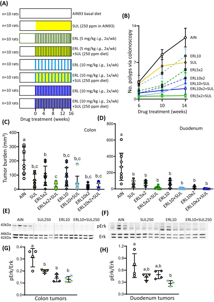
A previously reported clinical trial in familial adenomatous polyposis (FAP) patients treated with erlotinib plus sulindac (ERL + SUL) highlighted immune response/interferon-γ signaling as a key pathway. In this study, we combine intermittent low-dose ERL ± SUL treatment in the polyposis in rat colon (Pirc) model with mechanistic studies on tumor-associated immune modulation. At clinically relevant doses, short-term (16 weeks) and long-term (46 weeks) ERL ± SUL administration results in near-complete tumor suppression in Pirc colon and duodenum (p < 0.0001). We identify a low-dose threshold for significant antitumor activity in Pirc rats given SUL at 125 ppm in the diet plus ERL at 5 mg/kg body weight via twice-weekly oral gavage (SUL125 + ERL5 × 2). Longitudinal analyses show diminished expression of MHC class I and II genes in polyps larger than Grade 5, a novel finding in the Pirc model. Treatment with ERL ± SUL upregulates the corresponding MHC and immune-associated factors in a subset of Pirc colon polyps, Pirc tumor cell lines, murine colon carcinoma cells, and FAP patient-derived organoids, with Nlrc5 playing a critical role in this effect. Imaging mass cytometry reveals that SUL125 + ERL5 × 2 increases tumor-associated Cd4+ T cells by ~2.6-fold (p < 0.05), with no apparent effect on Cd8+ T cells. The treatment also increases tumor-associated Cd68+ cells (p < 0.05) and decreases Foxp3+ (p < 0.01) and Arg1+ (p < 0.05) cells. Thus, intermittent low-dose ERL + SUL treatment enhances tumor-associated MHC expression and remodels the immune cell niche toward a more permissive "helper" immune microenvironment. We conclude that early immune-interception strategies targeting interferon-γ signaling may benefit FAP patients at drug doses below the clinical standard of care.

Keywords: MHC; erlotinib; familial adenomatous polyposis (FAP); sulindac; tumor immunomodulation.


Conflict of interest statement

E. Vilar has consulting/advisory roles with Janssen Research, Recursion Pharma, Guardant Health (Sept 2022), Rising Tide Foundation (May 2023), Nouscom, Abbvie, and research support from Janssen Research. P.H. Brown is a stockholder in GeneTex (less than 1%), which is unrelated to this work. The other authors declare no potential conflicts of interest.

Figures

FIGURE 1
FIGURE 1
Short‐term intermittent low‐dose ERL ± SUL treatment suppressed tumor formation in the colon and duodenum of the Pirc model and reduced pErk expression. (A) Short‐term efficacy protocol involving 16‐week dosing of test agents. (B) Temporal tracking of antitumor response by colonoscopy (mean ± SE). (C, D) Scatter plots of antitumor activity in the colon and duodenum, respectively, at 16 weeks, with each animal represented by a single datapoint; horizontal line, median. (E, F) Representative immunoblots of pErk and Erk in Pirc colon and duodenum tumors, respectively, at 16 weeks. Each lane represents an individual polyp, and the data are representative of three or more independent experiments. In panels (G, H) immunoblots were quantified from Pirc colon and duodenum tumors, respectively, and groups sharing the same superscript letter were not significantly different by one way ANOVA (GraphPad Prism 9.5).
FIGURE 2
FIGURE 2
Long‐term intermittent low‐dose ERL ± SUL treatment suppressed Pirc colon and duodenum tumors. (A) Long‐term efficacy protocol involving 46‐week dosing of test agents. (B) Kaplan–Meier survival curves for rats up to 1 year of age. (C) Colonoscopic assessment of tumor burden following 29‐week dosing of test agents. In the scatter plots, each observable colon tumor was represented by a datapoint; horizontal line, median. Records also were kept according to tumor size Grade 1–5, with representative images provided. *p < 0.05 by Student's t‐test for each individual treatment group versus the corresponding AIN control. (D, E) Scatter plots of antitumor activity in the colon and duodenum of rats at 1 year, respectively, following 46‐week dosing of test agents, with each animal represented by a single datapoint. Groups sharing the same superscript letter were not significantly different by one‐way ANOVA.
FIGURE 3
FIGURE 3
Long‐term intermittent low‐dose SUL ± ERL treatment increased MHC gene expression in a sub‐population of Pirc tumors at 1 year and altered the immune cell niche. (A) Scatter plots of MHC class I and II gene expression assessed by RT‐qPCR and normalized to Gapdh; horizontal line, median. Data are representative of three or more independent experiments, with n = 9 colon tumors per treatment group. (B) IMC data were visualized as t‐SNE plots for selected markers of the immune cell niche in Pirc colon tumors at 1 year. (C) IMC data quantified (n = 3 tumors per group) revealed significant drug‐induced changes in the percentage of Cd4+, Cd68+, Foxp3+, and Arg1+ cells. In panels (A) and (C), groups with the same superscript letter were not significantly different by one‐way ANOVA.
FIGURE 4
FIGURE 4
Long‐term intermittent low‐dose SUL ± ERL treatment reduced Cd163 expression in Pirc colon and duodenum tumors. (A) Scatter plot of Cd163 gene expression in Pirc colon tumors at 1 year assessed by RT‐qPCR and normalized to Gapdh; horizontal line, median. Data are representative of three or more independent experiments (n = 9 tumors per group). Immunoblots of Cd163 expression in (B) Pirc colon and (C) Pirc duodenum tumors at 1 year, with β‐actin as loading control. Each lane represents a single tumor, and data are representative of three or more independent experiments. (D, E) Quantification of data in panels (B, C) respectively, by densitometry. In the scatter plots, groups sharing the same superscript letter were not significantly different by one‐way ANOVA.
FIGURE 5
FIGURE 5
Pirc tumors exhibit decreased MHC gene expression in large polyps, with reversibility demonstrated by ex vivo SUL ± ERL treatment. (A) Scatter plots of MHC class I and II gene expression assessed by RT‐qPCR and normalized to Gapdh; median, horizontal line. Data are representative of three or more independent experiments (n = 9 colon tumors per group). G1–G5 signifies polyp size (see Figure 2C), with occluding lesions being larger than 150 mm3 (>G5); Adj, adjacent. (B, C) Pirc colon tumor (PCT) and (D, E) mouse MC38‐OVA colon carcinoma cells incubated for 7 days with 0.75 mM ERL, 50 mM SUL, or ERL + SUL exhibited no significant changes in viability or live cell number compared to vehicle controls. No phenotypic alterations were observed after a 7‐day treatment with test agents (Figure S8). Immunoblots of (F), PCT and (G), MC38 cells after 7‐day treatment with SUL ± ERL, as in panels (B–E). MHC‐related proteins were normalized to β‐actin. Poly(ADP‐ribose) polymerase (PARP) served as an apoptosis marker. In PCT and MC38‐OVA cells, MHC‐I refers to rat RT‐1A and mouse H2‐d proteins, respectively. (H) Immunoassay‐based assessment of interleukin‐2 (Il‐2) secretion in a co‐culture assay, as reported, with IFN‐γ as the positive control. (I) Immunoblots of MC38‐OVA cells transfected with Nlrc5 siRNA (200 pmol) and treated concomitantly with ERL (0.75 μM) ± SUL (50 μM) for 7 days. β‐actin served as loading control. Scrambled siRNA served as negative control. (J) Working model for the non‐inducibility of MHC‐I pathway members following Nlrc5 KD (image created using software from BioRender.com, accessed on May 30, 2024). In panels A, B and D, groups sharing the same superscript letter were not significantly different by one‐way ANOVA.
FIGURE 6
FIGURE 6
The prototypical MHC class I‐dependent gene B2M is upregulated by ERL ± SUL treatment in FAP patient‐derived colon organoids. (A) Representative bright‐field microscopy images of FAP patient‐derived colon organoids at 4× magnification after 7‐day treatment with SUL, ERL, or ERL + SUL at the concentrations indicated. No marked phenotypic changes were observed or signs of overt toxicity. (B) Upregulation of B2M expression after treatment with test agents, as in panel (A), assessed by RT‐qPCR and normalized to GAPDH; horizontal line, median. Groups sharing the same superscript letter were not significantly different by one‐way ANOVA. (C) Working model for MHC class I pathway and its upregulation by ERL ± SUL in Pirc tumors and FAP patient‐derived organoids (image created using software from BioRender.com, accessed on January 26, 2024). IFN‐γ, interferon‐γ; IL‐2, interleukin‐2; GrB, granzyme B; Prf, perforin.

References

    1. Penny HL, Prestwood TR, Bhattacharya N, et al. Restoring retinoic acid attenuates intestinal inflammation and tumorigenesis in Apc Min/+ mice. Cancer Immunol Res. 2016;4:917‐926. - PMC - PubMed
    1. Lynch P. Chemoprevention of familial adenomatous polyposis. Fam Cancer. 2016;15:467‐475. - PubMed
    1. Kemp Bohan PM, Mankaney G, Vreeland TJ, et al. Chemoprevention in familial adenomatous polyposis: past, present and future. Fam Cancer. 2021;20:23‐33. - PMC - PubMed
    1. Samadder NJ, Neklason DW, Boucher KM, et al. Effect of sulindac and erlotinib vs placebo on duodenal neoplasia in familial adenomatous polyposis: a randomized clinical trial. Jama. 2016;315(12):1266‐1275. doi:10.1001/jama.2016.2522 - DOI - PMC - PubMed
    1. Samadder NJ, Kuwada SK, Boucher KM, et al. Association of sulindac and erlotinib vs placebo with colorectal neoplasia in familial adenomatous polyposis: secondary analysis of a randomized clinical trial. JAMA Oncol. 2018;4:671‐677. - PMC - PubMed

MeSH terms