

Amphipathic Antimicrobial Peptides Illuminate a Reciprocal Relationship Between Self-assembly and Cytolytic Activity
- PMID: 40073424
- PMCID: PMC12088898
- DOI: 10.1002/anie.202500040
Amphipathic Antimicrobial Peptides Illuminate a Reciprocal Relationship Between Self-assembly and Cytolytic Activity
Abstract
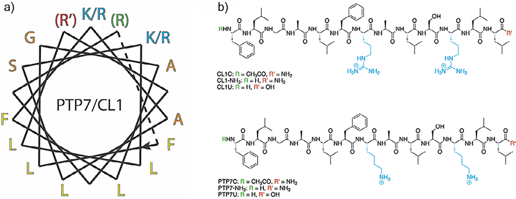
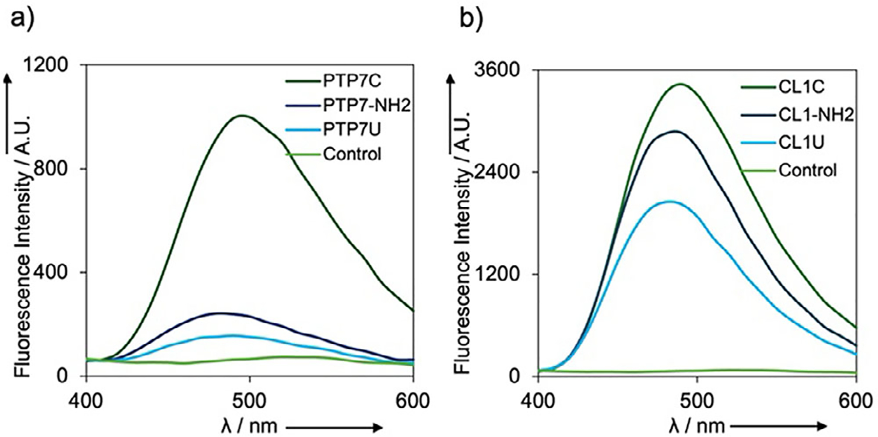
Amphipathic character, encoded within the polar sequence patterns of antimicrobial peptides, is a critical structural feature that influences membrane disruptive behavior. Similarly, polar sequence patterns induce self-assembly of amphipathic peptides, which results in the formation of ordered supramolecular structures. The relationship between self-assembly and membrane activity remains an open question of relevance for the development of effective antimicrobial peptides. Here, we report the structural investigation of a class of lytic peptides that self-assemble into filamentous nanomaterials. CryoEM analysis was employed to determine the structure of one of the filaments, which revealed that the peptides are self-assembled into a bilayer nanotube, in which the interaction between layers of amphipathic α-helices was mediated through hydrophobic interactions. The relative stability of the filament peptide assemblies depended on the influence of sequence modifications on the helical conformation. Antimicrobial assays indicated that cytolytic activity was associated with dynamic disassociation of the filamentous assemblies under the assay conditions. Structural modifications of the peptides that stabilized the filaments abrogated lytic activity. These results illuminate a reciprocal relationship between self-assembly and antimicrobial activity in this class of amphipathic peptides and that reversible assembly was critical for the observation of biological activity.
Keywords: Amphipathic sequence; Antimicrobial peptide; Polar pattern; Self‐assembly; cryoEM.
© 2025 Wiley‐VCH GmbH.
Conflict of interest statement
Conflict of Interests
The authors declare no conflict of interest.
Figures


References
MeSH terms
Substances
Grants and funding
- DBI-17265/National Science Foundation
- T32 AI106699/AI/NIAID NIH HHS/United States
- K99 GM138756/GM/NIGMS NIH HHS/United States
- U24 GM116790/GM/NIGMS NIH HHS/United States
- GM119426/NH/NIH HHS/United States
- R35 GM122510/GM/NIGMS NIH HHS/United States
- R35 GM119426/GM/NIGMS NIH HHS/United States
- AI106699/NH/NIH HHS/United States
- UL1 TR000454/TR/NCATS NIH HHS/United States
- S10 RR025067/RR/NCRR NIH HHS/United States
- GM138756/NH/NIH HHS/United States
- CHE-2108621/National Science Foundation
- G20 RR031199/RR/NCRR NIH HHS/United States
- R00 GM138756/GM/NIGMS NIH HHS/United States
- GM122510/NH/NIH HHS/United States
LinkOut - more resources
Full Text Sources
Medical
