. 2025 May 24;40(5):639-655.
doi: 10.1093/jbmr/zjaf040.

Heme metabolism mediates RANKL-induced osteoclastogenesis via mitochondrial oxidative phosphorylation

Affiliations

Heme metabolism mediates RANKL-induced osteoclastogenesis via mitochondrial oxidative phosphorylation

Heng Qiu et al. J Bone Miner Res. .

Abstract

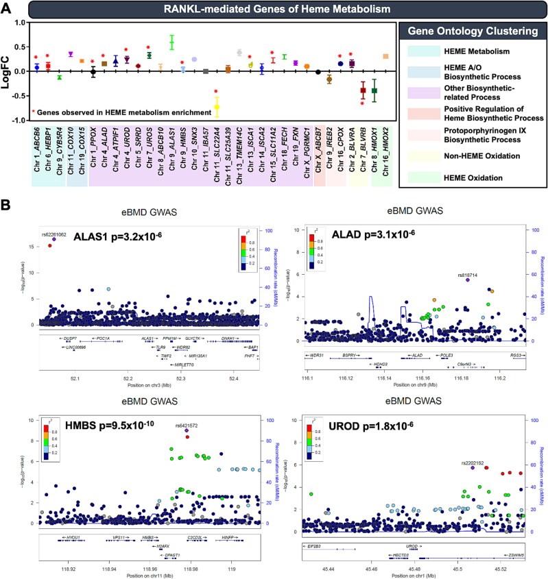
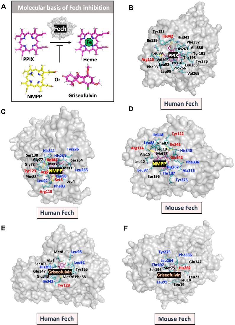
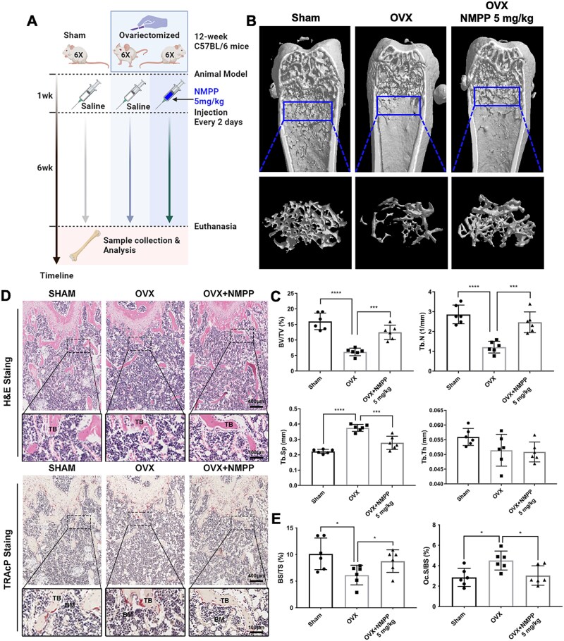
Bone undergoes life-long remodeling, in which disorders of bone remodeling could occur in many pathological conditions including osteoporosis. Understanding the cellular metabolism of osteoclasts (OCs) is key to developing new treatments for osteoporosis, a disease that affects over 200 million women worldwide per annum. We found that human OC differentiation from peripheral blood mononuclear cells derived from 8 female patients is featured with a distinct gene expression profile of mitochondrial biogenesis. Elevated mitochondrial membrane potential (MMP, Δψm) was also observed in receptor activator of NF-κB ligand (RANKL)-induced OCs. Interestingly, the gene pathways of heme synthesis and metabolism were activated upon RANKL stimulation, featured by transcriptomic profiling in murine cells at a single-cell resolution, which revealed a stepwise expression pattern of heme-related genes. The real-world human data also divulges potential links between heme-related genes and bone mineral density. Heme is known to have a role in the formation of functional mitochondrial complexes that regulate MMP. Disruption of heme biosynthesis via genetically silencing Ferrochelatase or a selective inhibitor, N-methyl Protoporphyrin IX (NMPP), demonstrated potent inhibition of OC differentiation, with a dose-dependent effect observed in NMPP treatment and a substantial efficacy even at a single dose. In vivo study further showed the protective effect of NMPP on ovariectomy-induced bone loss in female mice. Collectively, we found that RANKL-mediated signaling regulated mitochondrial formation and heme metabolism to synergistically support osteoclastogenesis. Inhibition of heme synthesis impaired OC formation and reversed excessive bone loss, representing a new therapeutic target for metabolic skeletal disorders.

Keywords: RANKL; heme; mitochondria; osteoclasts; osteoporosis.

Plain language summary

Bone remodeling is a lifelong process that can be disrupted in conditions like osteoporosis, a disease affecting over 200 million women globally per annum. Developing new treatments for osteoporosis requires understanding the cellular metabolism of osteoclasts (OCs), cells responsible for breaking down bone. In this study, we discovered that OC differentiation is characterized by increased mitochondrial biogenesis and elevated mitochondrial membrane potential (MMP). We also revealed that pathways involved in heme synthesis and metabolism were activated during this process, with real-world human data showing potential links between heme-related genes and bone mineral density. Heme is essential for forming functional mitochondrial complexes that regulate MMP and energy production, in which genetically knocking down heme catalyzing enzyme Ferrochelatase potently hampered receptor activator of NF-κB ligand (RANKL)-induced osteoclastogenesis. We found that a selective inhibitor of heme biosynthesis, N-methyl Protoporphyrin IX, effectively inhibited OC differentiation in a dose-dependent manner and protected against bone loss in estrogen-deficiency mice. These findings suggest that RANKL-mediated signaling regulates mitochondrial formation and heme metabolism to support osteoclastogenesis. Inhibiting heme synthesis could be a novel therapeutic target for treating metabolic skeletal disorders like osteoporosis.


Conflict of interest statement

None declared.

Figures

Graphical Abstract
Graphical Abstract
Figure 1
Figure 1
Transcriptome and GSEA analysis of gene expression in human OCs versus monocytes. (A) Brief illustration of transcriptome analysis of human OC differentiation. (B) TRAcP-positive human OCs with an enlarged image showing staining details. Scale bar = 200 μm. (C) The fold changes of mitochondrial markers during human OC differentiation. n = 8 pairs. (D) Fluorescence staining of JC-1 in RANKL-induced murine osteoclastogenesis. Increased J-aggregates suggests an elevated MMP (Δψ) induced by RANKL. Result was quantified by comparison of the fluorescence intensity of J-aggregates and monomers before and after 5-day RANKL treatment. Scale bar = 100 μm. n = 4. (E) GSEA enrichment plot in heme metabolism (MSigDB gene set: HALLMARK_HEME_METABOLISM). (F) Gene expression levels of essential enzymes for heme biosynthesis. (G) Heme content in murine BMMs treated without/with RANKL (n = 3). All data are displayed as mean ± SD. *p <.05, **p <.01, ***p <.001, and ****p <.0001 compared to PBMC or indicated otherwise.
Figure 2
Figure 2
RANKL regulates different biological processes of heme metabolism that are potentially associated with estimated bone mineral density (eBMD) by human GWAS. (A) The dot bar chart specifies the genes that modulate the biological processes of heme metabolism and their corresponding fold changes derived from our RNA sequencing dataset. n = 16/8 pairs. The bar graph is displayed as mean ± SD. (B) Regional association plots depicting eBMD GWAS results for genes encoding essential enzymes of heme biosynthesis. Genetic variants within 200 kb of each gene are plotted according to their chromosomal location (x-axis) and eBMD p-value (−log10) (y-axis). Variants are color-coded according to the degree of linkage disequilibrium (1000GP Nov 2014 EUR population) with the lead variant at each locus, with the recombination rate indicated by the blue line.
Figure 3
Figure 3
Deciphering RANKL-induced expression pattern of mitochondrial markers and heme-related genes in murine osteoclastogenesis at single-cell resolution. (A) The trajectory of OC differentiation was estimated by pseudotime analysis and mapped on a UMAP visualization. (B) Stepwise expression of mitochondrial markers during OC differentiation. (C) Dynamic and sequential expression of heme biosynthesis genes following the trajectory of osteoclastogenesis at single-cell resolution. (D) Quantification and statistical analysis of the expression pattern of mitochondrial markers and heme-related genes. All data are presented as mean ± SD. *p <.05, **p <.01, ***p <.001 and ****p <.0001 OP, OC and MC compared to MP; #p <.05, ##p <.01, and ####p <.0001 PC and MC compared to OP; &&&&p <.0001 MC compared to PC.
Figure 4
Figure 4
Knockdown of Fech gene disrupted OC differentiation. (A) Left panel: shRNA-mediated Fech knockdown significantly impaired RANKL-induced TRAcP-positive multinucleated cells from BMMs. Scale bar = 200 μm. Right panel: Statistical analysis of TRAcP staining. N = 3. All data are presented as mean ± SD. **p <.01 compared to sh-NC group. (B) rtPCR revealed that osteoclastic genes were profoundly downregulated following Fech knockdown during RANKL-mediated differentiation. n = 5. All data are presented as mean ± SD. ***p <.001 and ****p <.0001 compared to sh-NC group. (C) A consistent result is supported by western blotting, which shows decreased protein levels of osteoclastic markers in Fech deficient BMMs upon RANKL stimulation. (D) Statistical analysis of western blotting results. Each protein expression was normalized to β-actin and quantified by ImageJ based on gray intensity. N = 3. All data are presented by the mean ± SD. *p <.05, **p <.01, and ***p <.001 compared to sh-NC.
Figure 5
Figure 5
The molecular basis of heme biosynthesis inhibitors. (A) NMPP and Griseofulvin, an FDA-approved antifungus drug, were reported to inhibit heme biosynthesis via FECH, which catalyzes the terminal step in the biosynthesis of heme, converting protoporphyrin IX (PPIX) into heme B. (B) The crystal structure of FECH and PPIX bound (PDB: 3HCO) showing interactive residues and polar contacts (red color-labeled residues and yellow dash line) with PPIX. (C) and (D) Artificial intelligence model (chai discovery) was used to mimic NMPP-human/mouse FECH interactions. Red color highlights residues predicted to form polar contacts (yellow dash line) with NMPP and blue color labels interactive residues that also involve PPIX binding. (E) and (F) Chai discovery was used to predict Griseofulvin-human/mouse FECH interactions. Red color highlights residues potentially forming polar contacts (yellow dash line), and blue color highlights interactive residues that mediate PPIX binding.
Figure 6
Figure 6
In vitro therapeutic potentials of heme biosynthesis inhibitor NMPP. (A) NMPP suppressed osteoclastogenesis in a dose-dependent manner. Representative images are presented with TRAcP staining. Scale bar = 200 μm. (B) TRAcP-positive multinucleated cells were quantified in a bar chart by ImageJ. N = 5. (C) Heme content in BMMs, BMMs stimulated with RANKL, and BMMs treated with both RANKL and 5 μm NMPP. N = 3. Data are presented as mean ± SD. **p <.01 and ***p <.001 compared to negative control group or as indicated. (D) Podosome belt staining shows the dose-dependent inhibition of osteoclastogenesis induced by NMPP. Scale bar = 200 μm. (E) Time-course OC formation assay indicates that a single-dose treatment of NMPP at day 3 achieved a stronger inhibitory effect than those of day 1 and day 5 only. Left panel: Representative images of full-well TRAcP staining with enlarged regions of interest. Scale bar = 200 μm. Right panel: Quantification of full-well TRAcP-positive multinucleated cells. N = 4. All data are presented as mean ± SD. ****p <.0001 compared to positive ctrl. &&&p <.001 and &&&&p <.0001 compared to day3. (F) and (G). Heme inhibition by NMPP suppressed the activity of transcriptional factors AP-1 and NF-κB in a dose-dependent manner. (H) The fluorescence intensity of TMRE staining reflected the MMP in live cells. N = 5. All data are presented as mean ± SD. **p <.01, ***p <.001 and ****p <.0001 compared to positive ctrl group unless indicated otherwise.
Figure 7
Figure 7
In vivo treatment of NMPP alleviated OVX-induced bone loss. (A) The schematic diagram of in vivo experimental design to study the protective effect of NMPP on OVX mice. (B) Representative images of micro-CT, which show that NMPP evidently rescued the excessive bone loss caused by ovariectomy. (C) The quantitative analyses of micro-CT results, including BV/TV, Tb.N, Tb.Sp and Tb.Th. N = 6. (D) Representative images of HE and TRAcP staining for decalcified bone tissues collected from sham mice, OVX mice treated with or without 5 mg/kg NMPP, respectively. (E) Quantification of histological measurements, including BS/TS (%) and Oc.S/BS (%). All data are presented as mean ± SD. *p <.05, **p <.01, ***p <.001 and ****p <.0001 compared to OVX group unless indicated otherwise. Abbreviations: BM: Bone marrow; BS/TS, bone surface/total surface; BV/TV, bone volume/tissue volume; HE, hematoxylin and eosin; Oc.S/BS, osteoclast surface/bone surface; TB, trabecular bone; Tb.N, trabecular number; Tb.Sp, trabecular separation; Tb.Th, trabecular thickness; TRAcP, tartrate-resistant acid phosphatase.
Figure 8
Figure 8
Graphical abstract illustrates the main findings of this study. RANKL-RANK signaling enhanced heme metabolism partially through transcription factors NFATc1 and AP-1, in which the elevated heme activity was coupled with mitochondrial biogenesis to facilitate OXPHOS, producing ATPs that boosted OC formation. Inhibition of heme biosynthesis by NMPP exhibited a dose-dependent inhibitory effect in MMP and osteoclastogenesis, reversing the excessive bone loss caused by ovariectomy. This study proposes a novel therapeutic target for osteoporosis.

References

    1. Compston JE, McClung MR, Leslie WD. Osteoporosis. Lancet. 2019;393(10169):364–376. 10.1016/S0140-6736(18)32112-3 - DOI - PubMed
    1. Lorentzon M. Treating osteoporosis to prevent fractures: current concepts and future developments. J Intern Med. 2019;285(4):381–394. 10.1111/joim.12873 - DOI - PubMed
    1. Lemma S, Sboarina M, Porporato PE, et al. Energy metabolism in osteoclast formation and activity. Int J Biochem Cell Biol. 2016;79(2016):168–180. 10.1016/j.biocel.2016.08.034 - DOI - PubMed
    1. Da W, Tao L, Zhu Y. The role of osteoclast energy metabolism in the occurrence and development of osteoporosis. Front Endocrinol (Lausanne). 2021;12(2021):675385. 10.3389/fendo.2021.675385 - DOI - PMC - PubMed
    1. Kim JM, Jeong D, Kang HK, Jung SY, Kang SS, Min BM. Osteoclast precursors display dynamic metabolic shifts toward accelerated glucose metabolism at an early stage of RANKL-stimulated osteoclast differentiation. Cell Physiol Biochem. 2007;20(6):935–946. 10.1159/000110454 - DOI - PubMed