. 2025 Apr;26(4):567-581.
doi: 10.1038/s41590-025-02104-y. Epub 2025 Mar 12.

Tuft cell IL-17RB restrains IL-25 bioavailability and reveals context-dependent ILC2 hypoproliferation

Affiliations

Tuft cell IL-17RB restrains IL-25 bioavailability and reveals context-dependent ILC2 hypoproliferation

Xiaogang Feng et al. Nat Immunol. 2025 Apr.

Abstract

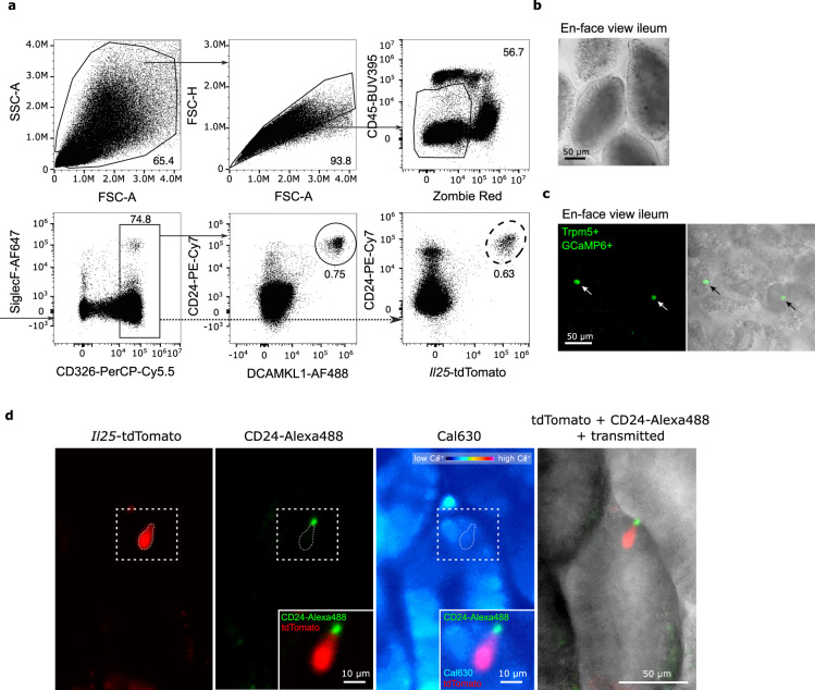
The tuft cell-group 2 innate lymphoid cell (ILC2) circuit orchestrates rapid type 2 responses upon detecting microbially derived succinate and luminal helminths. Our findings delineate key mechanistic steps involving IP3R2 engagement and Ca2+ flux, governing interleukin-25 (IL-25) production by tuft cells triggered by succinate detection. While IL-17RB has a pivotal intrinsic role in ILC2 activation, it exerts a regulatory function in tuft cells. Tuft cells exhibit constitutive Il25 expression, placing them in an anticipatory state that facilitates rapid production of IL-25 protein for ILC2 activation. Tuft cell IL-17RB is crucial for restraining IL-25 bioavailability, preventing excessive tonic ILC2 stimulation due to basal Il25 expression. Supraoptimal ILC2 stimulation by IL-25 resulting from tuft cell Il17rb deficiency or prolonged succinate exposure induces a state of hypoproliferation in ILC2s, also observed in chronic helminth infection. Our study offers critical insights into the regulatory dynamics of IL-25 in this circuit, highlighting the delicate tuning required for responses to diverse luminal states.


Conflict of interest statement

Competing interests: The authors declare no competing interests.

Figures

Fig. 1
Fig. 1. Succinate-induced IL-25 activates ILC2s in an ILC2-intrinsic, IL-17RB-dependent manner.
a, Expression of IL-17RB and KLRG1 by lineage-negative (Lin) cells. bd, Frequencies of IL-17RB+ (b), Ki-67+ (c) and IL-13 (Sm13)+ (d) ILC2s quantified by flow cytometry (n = 9–10 mice). e, Experimental scheme (top) and expression of Ki-67 and the IL-13 (Sm13) reporter by ILC2s (bottom). f,g, Frequencies of IL-13 (Sm13)+ (f) and Ki-67+ (g) ILC2s quantified by flow cytometry (n = 9–12 mice). h,i, Percentage of small intestinal IL-13 (Sm13)+ (h) and Ki-67+ (i) ILC2s quantified by flow cytometry from Il5R/R and Il5R/R; Il17rbfl/fl mice treated with succinate for 4 days (n = 8–9 mice). j, N. brasiliensis (N.b.) worm burden in the small intestine on day 9 after infection (n = 11 mice). Data are indicated as mean or mean ± s.e.m. (bd, fj). Data were analyzed using the Mann–Whitney test (bd, f, g, j) or a two-tailed unpaired t test (h, i). **P = 0.01–0.001; ***P = 0.001–0.0001; ****P < 0.0001.
Fig. 2
Fig. 2. Succinate-induced tuft cell activation depends on IP3R2-mediated cytosolic Ca2+ activity.
ac, Il25tdTomato reporter mice were treated with succinate or regular drinking water. The small intestine was analyzed on day 4. a, Il25tdTomato expression in CD24+EpCAM+ tuft cells determined by flow cytometry and compared to tuft cells from a reporter-negative mouse. b, Frequencies of Il25tdTomato+CD24+ tuft cells gated on EpCAM+CD45low/– cells. c, Percentage of Il25tdTomato+ cells (n = 6 mice). d, Proteins detected by liquid chromatography–mass spectrometry (LC–MS) in tuft cells, ranked by relative abundance. Tuft cell-specific marker proteins are labeled with their UniProt identifiers (n = 5 mice). ej, Tuft cell Ca2+ activity in Trpm5Cre; R26GCaMP6f mice. e, Tuft cells (green) in ex vivo scraped villus preparations; a GCaMP6f-expressing tuft cell (white box) is magnified. f, Reproducible Ca2+ response time courses of a tuft cell induced by 1 mM succinate. g, Ca2+ response time courses of a tuft cell to increasing succinate concentrations and no succinate control (n = 4–10 tuft cells, villus preparations from two to three mice). h, Dose–response curve of Ca2+ peak responses normalized to the maximum response of a tuft cell. i, Example traces of tuft cells stimulated with 1 mM succinate before (control) and after 10 μM CPA (blue). j, Peak Ca2+ responses from preparations as in h. Fpeak values in nine random tuft cells (villus preparations from three mice) are shown. k, Itpr1, Itpr2 and Itpr3 mRNA expression in tuft and nontuft cells from the small intestine (published dataset: Nadjsombati et al.; n = 3 mice). lo, Tuft cell Ca2+ activity with the Cal-630 indicator dye in villus preparations from Il25tdTomato Itpr2+/– and Itpr2–/– mice. l, Il25tdTomato-expressing tuft cells (red) after Cal-630 indicator loading. m, Percentage of succinate-responding tuft cells (n = 107–117 tuft cells from five mice per genotype; Mann–Whitney test). n,o, Examples of Ca2+ response time courses (n) and quantification of peak response (o) to 1 mM succinate and 60 mM KCl (positive control) in tuft cells from Il25tdTomato Itpr2+/– and Itpr2–/– mice (Fpeak values of 10–11 independent tuft cell measurements from three mice per genotype). Data were analyzed using the Mann–Whitney test (c, h, m, o), a paired t test (j) or a two-tailed unpaired t test (k). Box plots display the interquartile (25–75%) ranges, median (line) and mean (black square) values with whiskers indicating s.d. values. *P = 0.01–0.05; **P = 0.01–0.001; ***P = 0.001–0.0001; ****P < 0.0001; NS (no significance), P ≥ 0.05.
Fig. 3
Fig. 3. Succinate-elicited IL-25 production in tuft cells is triggered by IP3R2.
a,b, Frequencies of small intestinal IL-13 (Sm13)+ (a) and Ki-67+ (b) ILC2s quantified by flow cytometry from Il25tdTomato; Itpr2–/– and Il25tdTomato; Itpr2+/– littermate mice treated with succinate for 4 days (n = 7–8 mice). c, Flow cytometry analysis of Il25tdTomato (Flare25) reporter and CD24 expression, gated on EpCAM+ cells. d, Il25 mRNA expression in tuft and nontuft epithelial cells sorted by fluorescence-activated cell sorting (FACS) from the proximal small intestine (n = 6–7 mice; ND, not detected in at least one sample). e,f, Frequencies of IL-13 (Sm13)+ (e) and Ki-67+ (f) ILC2s quantified by flow cytometry from Il25tdTomato; Itpr2–/– and Il25tdTomato; Itpr2+/– mice treated with 1 μg rIL-25 on two consecutive days (n = 11–12 mice). g, Mice expressing the IL-13 (Sm13) reporter were treated with succinate for 4 days and injected with PBS or an anti-IL-17RB blocking antibody on days 0 and 2. h,i, Frequencies of IL-13 (Sm13)+ (h) and Ki-67+ (i) ILC2s quantified by flow cytometry (n = 5–6 mice). Data are indicated as mean or mean ± s.e.m (a, b, df, hi). Data were analyzed using a two-way analysis of variance (ANOVA) (a, b), a one-way ANOVA (h, i) or the Mann–Whitney test (e, f). *P = 0.01–0.05; **P = 0.01–0.001; ****P < 0.0001; NS, P ≥ 0.05.
Fig. 4
Fig. 4. Tuft cell IL-17RB restrains homeostatic circuit activation.
a, Expression of IL-17RB in small intestinal tuft cells from Vil1Cre; Il17rbfl/fl, Il17rbfl/fl and Il17rb–/– mice quantified by flow cytometry. b, Mean fluorescence intensity (MFI) of IL-17RB expression in tuft cells quantified by flow cytometry from Il25tdTomato reporter mice treated with or without succinate for 4 days (n = 7–8 mice). c, Il17rb mRNA expression in tuft and nontuft epithelial cells FACSorted from the proximal small intestine of Il25tdTomato reporter mice treated with or without succinate for 4 days (n = 6–7 mice). d, Representative immunofluorescence images of DCLK1 (yellow) in small intestines from naive Vil1Cre; Il17rbfl/fl and Il17rbfl/fl mice. e, Quantification of tuft cells by flow cytometry (n = 12–13 mice). f, Flow cytometry analysis of Ki-67 and IL-13 (Sm13) reporter expression by ILC2s. g,h, Frequencies of IL-13 (Sm13)+ (g) and Ki-67+ (h) ILC2s quantified by flow cytometry (n = 23–24 mice). i, Heatmaps showing the relative expression of ILC2 signature genes (left) and genes associated with ILC2 activation (right), generated from bulk RNA sequencing of FACSorted ILC2s (CD45+LinKLRG1+) from the small intestines of Vil1Cre; Il17rbfl/fl and Il17rbfl/fl mice (n = 4–5 samples). j,k, Frequencies of small intestinal IL-13 (Sm13)+ (j) and Ki-67+ (k) ILC2s quantified by flow cytometry from Vil1CreERT2; Il17rbfl/fl and Il17rbfl/fl mice 1 or 6 weeks after tamoxifen treatment (n = 9–14 mice). ln, Frequencies of small intestinal IL-13 (Sm13)+ ILC2s (l), Ki-67+ ILC2s (m) and tuft cells (n) quantified by flow cytometry from Il25iCre/+; Il17rbfl/fl and Il17rbfl/fl mice (n = 8 mice). Data are indicated as median (b, c) or mean ± s.e.m (e, g, h, j, k, ln). Data were analyzed using the Mann–Whitney test (b, c, e, g, h, j, k, ln). *P = 0.01–0.05; **P = 0.01–0.001; ***P = 0.001–0.0001; ****P < 0.0001; NS, P ≥ 0.05.
Fig. 5
Fig. 5. Tuft cell-intrinsic IL-17RB controls microbiota-independent ILC2 activation in young mice.
a, Flow cytometry analysis of small intestinal tuft cells from Vil1Cre; Il17rbfl/fl and Il17rbfl/fl mice, gated as DCLK1+CD24+ cells of EpCAM+ cells. b,c, Frequencies of IL-13 (Sm13)+ ILC2s at 1 week (b) or 3 weeks (c) of age, quantified by flow cytometry (n = 6–9 mice). d,e, Expression (d) and quantification (e) of KLRG1 in ILC2s, analyzed by flow cytometry (n = 5–7 mice). f, Flow cytometry analysis of IL-13 (Sm13) reporter expression by ILC2s from 3-week-old Il25iCre/+; Il17rbfl/fl and Il17rbfl/fl mice. g, Quantification of IL-13 (Sm13)+ ILC2s (n = 6–7 mice). h,i, Flow cytometry analysis of KLRG1 expression by ILC2s (h) and quantification of the KLRG1 MFI (i) (n = 6–7 mice). j, Flow cytometry analysis of Ki-67 and Sm13 reporter expression by ILC2s from 3-week-old Vil1Cre; Il17rbfl/fl and Il17rbfl/fl mice, treated before and after birth with broad-spectrum antibiotics (ABX: metronidazole, neomycin sulfate, vancomycin and ampicillin) in drinking water. k, Quantification of IL-13 (Sm13)+ ILC2s (n = 4–8 mice). l,m, Flow cytometry analysis of KLRG1 expression by ILC2s (l) and quantification of the KLRG1 MFI (m) (n = 4–8 mice). Data were analyzed using the Mann–Whitney test and represented as mean ± s.e.m (b, c, e, g, i, k, m). **P = 0.01–0.001; ****P < 0.0001; NS, P ≥ 0.05.
Fig. 6
Fig. 6. Tuft cell-intrinsic IL-17RB regulates tonic IL-25 bioavailability.
a, Flow cytometry analysis of Ki-67 and Sm13 expression by small intestinal ILC2s from 3-week-old mice of the indicated genotypes. b, Quantification of IL-13 (Sm13)+ ILC2s (n = 6–9 mice). c, Analysis of KLRG1 expression by ILC2s. d, Quantification of KLRG1 MFI in ILC2s (n = 6–9 mice). e, Expression of tdTomato and CD24 in CD45low/–EpCAM+ cells determined by flow cytometry analysis from 3-week-old Vil1Cre; Il25fl/fl and Il25fl/fl mice, which also encode the Il25tdTomato reporter, and compared to that in a reporter-negative mouse. f,g, Frequencies of IL-13 (Sm13)+ (f) and Ki-67+ (g) ILC2s (n = 10–11 mice). h, Quantification of the KLRG1 MFI in ILC2s (n = 10–11 mice). i, Quantification of tuft cells by flow cytometry (n = 10–11 mice). j, Experimental scheme. k, Flow cytometry analysis of Ki-67 and Sm13 reporter expression by ILC2s from young Vil1Cre; Il17rbfl/fl and Il17rbfl/fl mice after five injections of PBS or an anti-IL-17RB blocking antibody. l, Quantification of IL-13 (Sm13)+ ILC2s (n = 9–11 mice). Data are indicated as mean or mean ± s.e.m (b, d, f, g, i, l). Data were analyzed using an ordinary one-way ANOVA (b, d, l) or the Mann–Whitney test (f, g, h, i). *P = 0.01–0.05; **P = 0.01–0.001; ****P < 0.0001; NS, P ≥ 0.05.
Fig. 7
Fig. 7. Prolonged activation by IL-25 induces a hypoproliferative state in ILC2s.
a,b, Percentage of IL-13 (Sm13)+ (a) and Ki-67+ (b) ILC2s in the small intestine quantified by flow cytometry from adult Vil1Cre; Il17rbfl/fl and Il17rbfl/fl littermate mice treated with succinate for 4 days (n = 6–13 mice). c,d, Percentage of IL-13 (Sm13)+ (c) and Ki-67+ (d) ILC2s in the small intestine quantified by flow cytometry from adult Vil1Cre; Il17rbfl/fl and Il17rbfl/fl littermate mice treated with 1 μg rIL-25 on two consecutive days (n = 9–12 mice). e, Flow cytometry analysis of Ki-67 and IL-13 (Sm13) reporter expression by ILC2s from IL-13 (Sm13) reporter mice treated with succinate for the indicated amount of time. f,g, Frequencies of IL-13 (Sm13)+ (f) and Ki-67+ (g) ILC2s (n = 8–10 mice; D, days; W, weeks). h, Percentage of DCLK1+CD24+ tuft cells among epithelial cells (n = 8–10 mice). i, Representative histograms displaying KLRG1 expression by ILC2s. j, Mice were treated with succinate for 4 weeks, followed by 2 weeks of regular drinking water and another 4 days of succinate treatment, and the small intestine was analyzed. k,l, Frequencies of Ki-67+ ILC2s (k) and MFI of IL-17RB in ILC2s (l) analyzed by flow cytometry (n = 6–9 mice). Data are indicated as mean ± s.e.m (ad, fh) or median (k, l). Data were analyzed using a two-way ANOVA (ad) or an ordinary one-way ANOVA (k, l). **P = 0.01–0.001; ***P = 0.001–0.0001; ****P < 0.0001; NS, P ≥ 0.05.
Fig. 8
Fig. 8. Stimulation of a nonredundant signaling pathway restores proliferative capacity in ILC2s.
a, Schematic of the signaling pathway engaged by the hM3Dq DREADD agonist CNO. b, KLRG1 and hM3Dq DREADD (HA-tag) in ILC2s, gated on CD45+Lin cells from Il5R/R and Il5R/R; TgGq-DREADD mice. c, Quantified percentage of hM3Dq DREADD+ ILC2s (n = 4–5 mice). d, Schematic showing 30-day succinate treatment with CNO injections on two consecutive days before analysis; used in e and f. e,f, Frequencies of small intestinal IL-13 (Sm13)+ (e) and Ki-67+ (f) ILC2s from Il5R/R and Il5R/R; TgGq-DREADD mice quantified by flow cytometry (n = 9–10 mice). g, IL-13 (Sm13) reporter mice were treated with succinate for 4 weeks, followed by NMU injections on two consecutive days. The frequencies of Ki-67+ ILC2s were analyzed by flow cytometry (n = 8–9 mice). h,i, Percentages of IL-13 (Sm13)+ (h) and Ki-67+ (i) small intestinal ILC2s analyzed by flow cytometry from Vil1Cre; Il17rbfl/fl and Il17rbfl/fl mice treated with NMU for two consecutive days (n = 7–14 mice). jl, YRS reporter mice infected with H. polygyrus were analyzed at the indicated time points. The frequencies of small intestinal IL-13 (Sm13)+ ILC2s (j), quantification of tuft cells (k) and frequencies of Ki-67+ ILC2s (l) were analyzed by flow cytometry (n = 6–7 mice). m, Il5R/R and Il5R/R; TgGq-DREADD mice were infected with third-stage larvae (L3) of H. polygyrus. Worms were cleared with pyrantel pamoate. Two weeks later, mice were treated with succinate water for 4 days and injected with CNO on two consecutive days before analysis. Control groups consisted of Il5R/R mice also treated with CNO and Il5R/R; TgGq-DREADD mice treated with PBS. n, Frequencies of Ki-67+ ILC2s in the small intestinal lamina propria analyzed by flow cytometry (n = 8–9 mice). Data are indicated as mean (cg, n) or mean ± s.e.m (hi). Data were analyzed using a two-tailed unpaired t test (c, e, f) or an ordinary one-way ANOVA (gl, n). *P = 0.01–0.05; **P = 0.01–0.001; ***P = 0.001–0.0001; ****P < 0.0001; NS, P ≥ 0.05.
Extended Data Fig. 1
Extended Data Fig. 1. ILC2-intrinsic requirement for IL-17RB in homeostatic and succinate-induced ILC2 activation.
a, Flow cytometry gating of small intestinal ILC2s, throughout the paper defined as either Gata3+KLRG1+ (fixed panel, filled line), or KLRG1+ (live panel, dashed line). b, Expression of IL-5 and IL-17RB by SI ILC2 cells from naïve Il5R/R;Il17rbfl/+, Il5R/R;Il17rbfl/fl, and Il5+/+;Il17rb–/– mice. Il5R allele: Il5tdTomato-Cre, known as “Red5”. c, Red5 or IL-17RB expression by CD3+CD4+ cells, gated as in a). d,e, Frequencies of IL-17RB+ (d) and Ki-67+ (e) ILC2s were quantified by flow cytometry (n = 7–12 mice). f, Expression of KLRG1 and Arg1YFP, IL-5 (Red5), and IL-13 (Sm13) reporters by Lin cells. g,h, Frequencies of IL-13 (Sm13)+ (g) and Ki-67+ (h) ILC2s were quantified by flow cytometry from Il5R/+;Il17rbfl/+, Il5R/+;Il17rbfl/fl and Il17rb–/– mice which were treated with succinate for 4 days (n = 7–14 mice). i, Frequencies of IL-17RB+ ILC2s were quantified by flow cytometry from Il5R/R and Il5R/R;Il17rbfl/fl mice treated with succinate for 4 days (n = 8–9 mice). Data mean (i) or mean ± s.e.m (d,e,g,h) is indicated. Data was analyzed using Ordinary one-way ANOVA (d,e,g,h) or two-tailed unpaired t test (i). *P 0.01–0.05; **, P 0.01–0.001; ***, P 0.001–0.0001;****, P < 0.0001; ns, P ≥ 0.05.
Extended Data Fig. 2
Extended Data Fig. 2. Visualization of Il25 expression and Ca2+ signaling in tuft cells.
a, Flow cytometry gating of small intestinal tuft cells, throughout the paper defined as either DCLK1+CD24+ (fixed panel, filled line), or Il25tdTomato+CD24+ double positive (live panel, dashed line). b, Tuft cell Ca2+ activity of the ileum was imaged with an ex vivo whole mount (en-face view of the ileum) preparation from Trpm5Cre;R26GCaMP6f mice. c, Tuft cells (green) can be identified based on Trpm5 promoter-dependent expression of GCaMP6f (arrows). d, Tuft cell Ca2+ activity of the ileum was imaged with an ex vivo whole mount preparation from Il25tdTomato (Flare25) mice using a Cal630 Ca2+ indicator dye. Tuft cells (white box) can be identified based on their distinct Il25 (red) and CD24 (green) (arrows).
Extended Data Fig. 3
Extended Data Fig. 3. Succinate-induced ILC2 activation is abolished in Itpr2–/– mice.
a, Flow cytometry analysis of Ki-67 and IL-13 (Sm13)-reporter expression by ILC2s from Itpr2+/– and Itpr2+/– mice treated for 4 days with succinate. b-d, The MFI of IL-13 (Sm13) (b) and Ki-67 (c) expression in ILC2s, and of the Il25tdTomato reporter in tuft cells (d) was quantified (n = 7–8 mice). e, Frequency of Il25tdTomato+ cells (n = 7–8 mice). Data median is indicated (b-e). Data was analyzed using two-way ANOVA(b-e). **, P 0.01–0.001; ***, P 0.001–0.0001; ****, P < 0.0001; ns, P ≥ 0.05.
Extended Data Fig. 4
Extended Data Fig. 4. Tuft cell IL-17RB restrains homeostatic circuit activation.
a, GFP reporter signal in tuft cells analyzed by flow cytometry of SI organoids from NF-κBGFP activity reporter mice treated with indicated cytokines for 24 h. b, Frequency of NF-kBGFP+ tuft cells (n = 1–2 technical replicates on 2 biological replicates). c, Tuft cell proteomics analysis (n = 3–5 mice). Volcano plot showing differentially expressed proteins in tuft cells from IL-25 treated mice compared to controls. Each point represents a detected protein. Proteins with significant expression changes are labelled with their UniProt identifiers. d, IF of MUC2 in SI (scale bars, 100 μm). e, Muc2 area relative to DAPI area per imaging field (n = 8 mice). f, SI ILC2s numbers (n = 14–15 mice). g, Expression of IL-13 (Sm13) and Arg1YFP reporters or IL-17RB by CD45+Lin cells. h, Ki-67 and IL-13 (Sm13)-reporter expression by lung ILC2s. i,j, Frequencies of IL-13 (Sm13)+ (i) and Ki-67+ (j) ILC2s, quantified by flow cytometry (n = 10 mice). k,l, SI length (k) and body weight (l) of the indicated mice (n = 6–9 mice). m, Day 10 SI N. brasiliensis worm burden (n = 5 mice). n, Fecal egg burden on day 8 and 28 post H. polygyrus infection (n = 19 mice). o, Worm burden 28 days after infection (n = 10–13 mice). p, IL-17RB MFI of ILC2s of Vil1CreERT2;Il17rbfl/fl mice, 6 weeks post tamoxifen (n = 8–9 mice). q, Il25 mRNA expression in tuft cells from Vil1CreERT2;Il17rbfl/fl and Il17rbfl/fl mice 1 week post tamoxifen (n = 8–9 mice). r, Frequencies of IL-17RB+ tuft cells in the indicated mice by flow cytometry (n = 3–5 mice). Data median (b,m-p) or mean ± s.e.m (e,f,i-l,q,r) is indicated. Data analyzed using Ordinary one-way ANOVA(b,r) or Mann-Whitney test (e,f,i-q). *P 0.01–0.05; **, P 0.01–0.001; ***, P 0.001–0.0001;****, P < 0.0001; ns, P ≥ 0.05.
Extended Data Fig. 5
Extended Data Fig. 5. Elevated tonic ILC2 stimulation in Vil1Cre;Il17rbfl/fl mice is independent of Il17rc.
a, Il25tdTomato MFI of EpCAM+ CD24+SiglecF+ tuft cells were determined by flow cytometry from 3-week-old Vil1Cre;Il25fl/fl and Il25fl/fl mice which also encode the Il25tdTomato reporter, and compared to that in a reporter-negative mouse (n = 4–10 mice). b, Representative immunofluorescence image of DCLK1 (green) in 2D organoids from mice of the indicated genotypes (scale bars, 200 μm). c,IL-25 production in supernatants of 2D organoids stimulated with DMSO, 1 µg/mL ionomycin, or 50 mM dibasic sodium succinate hexahydrate for 30 min. The dashed line indicates the limit of detection (n = 6 samples from 6 mice). d,e, Enumeration of IL-13 (Sm13)+ ILC2 percentage (d) and quantification of the KLRG1 MFI in ILC2s (e) (n = 4–8 mice). Data are represented as mean ± s.e.m (a,c-e). Data was analyzed using Ordinary one-way ANOVA (a,c-e). **, P 0.01–0.001; ***, P 0.001–0.0001;****, P < 0.0001; ns, P ≥ 0.05.
Extended Data Fig. 6
Extended Data Fig. 6. Prolonged activation by IL-25 induces a hypoproliferative state in ILC2s.
a,b, The MFI of Il25tdTomato reporter in tuft cells (a) and the percentages of Il25tdTomato+ cells (b) were quantified by flow cytometry. The SI was analyzed from mice treated with succinate for 4 weeks, followed by 2 weeks of regular drinking water, and another 4 days of succinate treatment, according to the schematic in (Fig. 7j), (n = 6–9 mice). c, The number of SI ILC2s was quantified from IL-13 (Sm13)-reporter mice treated with succinate for 4 weeks, followed by neuromedin U (NMU) injections on two consecutive days. (n = 8–9 mice). Data are represented as median (a-c). Data were analyzed using Ordinary one-way ANOVA(a-c). *, P 0.01–0.05; **, P 0.01–0.001; ***, P 0.001–0.0001; ns, P ≥ 0.05.

Update of

References

    1. Schneider, C. Tuft cell integration of luminal states and interaction modules in tissues. Pflugers Arch.473, 1713–1722 (2021). - PMC - PubMed
    1. Strine, M. S. & Wilen, C. B. Tuft cells are key mediators of interkingdom interactions at mucosal barrier surfaces. PLoS Pathog.18, e1010318 (2022). - PMC - PubMed
    1. von Moltke, J., Ji, M., Liang, H.-E. & Locksley, R. M. Tuft-cell-derived IL-25 regulates an intestinal ILC2–epithelial response circuit. Nature529, 221–225 (2016). - PMC - PubMed
    1. Howitt, M. R. et al. Tuft cells, taste-chemosensory cells, orchestrate parasite type 2 immunity in the gut. Science351, 1329–1333 (2016). - PMC - PubMed
    1. Gerbe, F. & Jay, P. Intestinal tuft cells: epithelial sentinels linking luminal cues to the immune system. Mucosal Immunol.9, 1353–1359 (2016). - PubMed

LinkOut - more resources