. 2025 Mar 12;16(1):170.
doi: 10.1038/s41419-025-07481-8.

Both direct and indirect suppression of MCL1 synergizes with BCLXL inhibition in preclinical models of gastric cancer

Affiliations

Both direct and indirect suppression of MCL1 synergizes with BCLXL inhibition in preclinical models of gastric cancer

Li-Ping Zhang et al. Cell Death Dis. .

Abstract

Despite the progress of treatment in gastric cancer (GC), the overall outcomes remain poor in patients with advanced diseases, underscoring the urgency to develop more effective treatment strategies. BH3-mimetic drugs, which inhibit the pro-survival BCL2 family proteins, have demonstrated great therapeutic potential in cancer therapy. Although previous studies have implicated a role of targeting the cell survival pathway in GC, the contribution of different pro-survival BCL2 family proteins in promoting survival and mediating resistance to current standard therapies in GC remains unclear. A systematic study to elucidate the hierarchy of these proteins using clinically more relevant GC models is essential to identify the most effective therapeutic target(s) and rational combination strategies for improving GC therapy. Here, we provide evidence from both in vitro and in vivo studies using a broad panel of GC cell lines, tumoroids, and xenograft models to demonstrate that BCLXL and MCL1, but not other pro-survival BCL2 family proteins, are crucial for GC cells survival. While small molecular inhibitors of BCLXL or MCL1 exhibited some single-agent activity, their combination sufficed to cause maximum killing. However, due to the unsolved cardiotoxicity associated with direct MCL1 inhibitors, finding combinations of agents that indirectly target MCL1 and enable the reduction of doses of BCLXL inhibitors while maintaining their anti-neoplastic effects is potentially a feasible approach for the further development of these compounds. Importantly, inhibiting BCLXL synergized significantly with anti-mitotic and HER2-targeting drugs, leading to enhanced anti-tumour activity with tolerable toxicity in preclinical GC models. Mechanistically, anti-mitotic chemotherapies induced MCL1 degradation via the ubiquitin-proteasome pathway mainly through FBXW7, whereas HER2-targeting drugs suppressed MCL1 transcription via the STAT3/SRF axis. Moreover, co-targeting STAT3 and BCLXL also exhibited synergistic killing, extending beyond HER2-amplified GC. Collectively, our results provide mechanistic rationale and pre-clinical evidence for co-targeting BCLXL and MCL1 (both directly and indirectly) in GC. (i) Gastric cancer cells rely on BCLXL and, to a lesser degree, on MCL1 for survival. The dual inhibition of BCLXL and MCL1 with small molecular inhibitors acts synergistically to kill GC cells, regardless of their TCGA molecular subtypes or the presence of poor prognostic markers. While the effect of S63845 is mediated by both BAX and BAK in most cases, BAX, rather than BAK, acts as the primary mediator of BCLXLi in GC cells. (ii) Inhibiting BCLXL significantly synergizes with anti-mitotic and HER2-targeting drugs, leading to enhanced anti-tumour activity with tolerable toxicity in preclinical GC models. Mechanistically, anti-mitotic chemotherapies induce MCL1 degradation via the ubiquitin-proteasome pathway mainly through FBXW7, whereas HER2-targeting drugs suppress MCL1 transcription via the STAT3/SRF axis. The combination of the STAT3 inhibitor and BCLXL inhibitor also exhibits synergistic killing, extending beyond HER2-amplified GC.


Conflict of interest statement

Competing interests: J.N.G. is a former employee of the Walter and Eliza Hall Institute of Medical Research, who receives milestones and royalty payments related to venetoclax. All the other authors declare no conflicts of interest. Ethics approval and consent to participate: All methods were performed in accordance with the relevant guidelines and regulations. Fresh tumor tissues were collected from GC patients during surgery after written informed consent was obtained. The study was performed with the approval of the National Cancer Center/National Clinical Research Center for Cancer/Cancer Hospital (17-156/1412) and the Institute of Laboratory Animal Sciences (GJN22002), Chinese Academy of Medical Sciences and Peking Union Medical College. Animal study was approved by the Institutional Animal Care and Use Committee of the Institute of Laboratory Animal Sciences, Chinese Academy of Medical Sciences, and Peking Union Medical College (IACUC 21001).

Figures

None
(i) Gastric cancer cells rely on BCLXL and, to a lesser degree, on MCL1 for survival. The dual inhibition of BCLXL and MCL1 with small molecular inhibitors acts synergistically to kill GC cells, regardless of their TCGA molecular subtypes or the presence of poor prognostic markers. While the effect of S63845 is mediated by both BAX and BAK in most cases, BAX, rather than BAK, acts as the primary mediator of BCLXLi in GC cells. (ii) Inhibiting BCLXL significantly synergizes with anti-mitotic and HER2-targeting drugs, leading to enhanced anti-tumour activity with tolerable toxicity in preclinical GC models. Mechanistically, anti-mitotic chemotherapies induce MCL1 degradation via the ubiquitin-proteasome pathway mainly through FBXW7, whereas HER2-targeting drugs suppress MCL1 transcription via the STAT3/SRF axis. The combination of the STAT3 inhibitor and BCLXL inhibitor also exhibits synergistic killing, extending beyond HER2-amplified GC.
Fig. 1
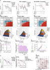
Fig. 1. A subset of human GC cell lines is susceptible to single inhibition of BCLXL or MCL1.
A Summary of the BH3-mimetic compounds used in this study. B Sensitivity of human GC cell lines to BH3-mimetic compounds. The sensitivity of 16 GC cell lines representing the four TCGA molecular subtypes to the indicated BH3-mimetics was determined after culturing in 0–10 μM of the drugs for 24 h. A discrete heat map representation of the mean IC50s was shown. C Genetic deleting BCLXL induces cell death in 23132/87 cells. The viability of 23132/87 cells 72 h after addition of doxocycline (DOX) to induce the expression of sgRNAs to target BCL2, BCLXL, BCLW, MCL1, BCL2A1 or BCLB was determined with CellTiter-Glo assays. 2 sgRNAs were tested for each gene. Unpaired Student’s t-test was used for statistical significance. D SNU-16 cells lose their viability when MCL1 is genetically ablated. Similar experiments to those in (C) were performed in SNU-16 cells. Anti-tumor activity of BCLXLi and MCL1i in vivo. BALB/c nude mice were inoculated s.c. with NCI-N87 (E) or SNU-668 (F) cells and treatment commenced 1 week later with BCLXLi (p.o.) or MCL1i (i.v.) as indicated. Tumor sizes were monitored every 3 days. Data shown represents the mean tumor volumes ± SD of 5 mice (E) and 7 mice (F) in each group. Statistical significance was calculated using the Two-way ANOVA analysis. G, H Correlation analysis of BCLXLi or MCL1i sensitivity and recurrent poor-risk genetic alterations in GC. All GC lines used in the study were ranked by increasing IC50s to BCLXLi (G) or MCL1i (H) treatment. The recurrent genetic alterations associated with poor prognosis in these cells were derived from the TCGA PanCancer Atlas study using the cBioPortal. Wilcoxon rank sum test was performed to determine the correlation between drug sensitivity and different genetic alterations. I Expression of BCL2 family proteins in the panel GC lines studied. J Spearman’s correlation analysis of BCL2 family protein expression and response to MCL1i and BCLXLi treatments. Spearman’s coefficient values with P value < 0.05 were shown in the plot. K Heatmap representation showing the impact of deleting BAX, BAK, or both on MCL1i and BCLXLi sensitivity. The mean IC50s ± SD of GC cell lines expressing sgRNAs targeting BAX, BAK or both to the indicated BH3-mimetic drugs were shown. Cell viability was determined using the CellTiter-Glo assay; data in (B–D) and (K) represent the means ± SD of ≥3 independent experiments; blots in (I) are representatives of 2 independent experiments. P values < 0.05 were considered significant. *P < 0.05, **P < 0.01, ***P < 0.001.
Fig. 2
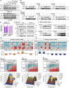
Fig. 2. Dual inhibition of BCLXL and MCL1 act synergistically to kill both GC cell lines and patient-derived organoids.
A Sensitivity of GC cell lines to different combinations of BH3-mimetic drugs. Similar experiments to those in Fig. 1B were performed with different combinations of BH3-mimetic drugs in equimolar concentrations (1:1 or 1:1:1). The responses of BCLXLi-sensitive, MCL1i-sensitive or MCL1i/BCLXLi-insensitive GC lines to the indicated treatments were summarized. Paired Student’s t-test was used to determine statistical significance. Synergistic killing by dual inhibition of BCLXL and MCL1. The viability of SNU-719 (B), 23132/87 (C) and HGC-27 (D) cells 24 h after treatment with increasing concentrations of indicated BH3-mimetic drugs was determined. BLISS scores were calculated and BLISS values > 0.0 indicate synergy between the two drugs at indicated concentrations. E BAX/BAK dependent killing of combining BCLXLi and MCL1i. The viability of WT and BAX/BAK deficient subclones of HGC-27 cells 24 h after treatment with the combination of BCLXLi and MCL1i (0–10 μM, 1:1) was determined. F Simultaneous deletion of BCLXL and MCL1 induces cell death in HGC-27 cells. Similar experiments to those in Fig. 1C, D were performed in HGC-27 cells. G Loss of BCLXL enhances MCL1 inhibition in vivo. HGC-27 cells inducibly expressing sgBCLXL or the sgRNA empty vector were subcutaneously inoculated into the BALB/c nude mice and treatment commenced 1 week later with doxycycline food alone or together with MCL1i. Tumor sizes were monitored every 3 days. Data shown represents the mean tumor volumes ±SD of 6 mice in each group. H Sensitivity of patient-derived organoids to the BH3-mimetic treatment. Established tumoroids were dissociated and seeded as single cells, grown into organoids over 7–9 days and treated with the indicated BH3-mimetic drugs, alone or in equimolar combinations (1:1 or 1:1:1). Cell viability was determined 24 h later using CellTitre-Glo 3D assays. Paired Student’s t-test was used to determine statistical significance. I Representative dose response curves of organoid #6 shown in (H). J The response of organoid #6 to different BH3-mimetic treatments was determined by PI (red) staining. Representative images at 72 h post-treatment were shown. Cell viability was determined using the CellTiter Glo or CellTiter Glo 3D assay; data in (A–F), (H) and (I) represent the means ± SD of ≥3 independent experiments; data in (J) are representatives of 2 independent experiments.
Fig. 3
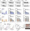
Fig. 3. Anti-mitotic chemotherapies induce MCL1 degradation and synergize with BCLXL inhibitor to kill GC cells.
A Effect of different chemotherapies on MCL1, BCLXL and BCL2 expression. SNU-216 and 23132/87 cells were treated with DMSO (Con) or 10 μM of the indicated chemotherapies for 24 h and the protein levels of MCL1, BCL2, BCLXL were determined by Western blotting. Ubiquitin-mediated protein degradation of MCL1 upon treatment with anti-mitotic drugs. SNU-216 (B) and 23132/87 cells (C) were treated with anti-mitotic drugs alone (10 μM) or together with inhibitors of proteasome (MG132, 5 μM) or lysosome (CQ, 5 μM) for 24 h and the protein levels of MCL1 were determined by Western blotting. D Effect of anti-mitotic drugs on MCL1 mRNA level. SNU-216 and 23132/87 cells were treated with indicated drugs (10 μM) for 24 h and the mRNA levels of MCL1 were determined by RT-qPCR. E Role of FBXW7 in mediating the degradation of MCL1 by anti-mitotic drugs. SNU-216 cells transduced with sgRNA targeting FBXW7 or the sgRNA empty vector were treated with anti-mitotic drugs (10 μM) for the indicated time periods. The protein levels of MCL1 were determined by Western blotting. F Effect of anti-mitotic drugs on MCL1 degradation in FBXW7-mutant cells. FBXW7-mutant MKN1 (R465C) cells were treated with anti-mitotic drugs alone (10 μM) or together with MG132 (5 μM) for the indicated time points and the protein levels of MCL1 were determined by Western blotting. G, H In vitro activity of BCLXLi or MCL1i in combination with different chemotherapies. The responses of MCL1i-sensitive (SNU-668, SNU-16, HGC-27), BCLXLi-sensitive (SNU-216, 23132/87, NCI-N87) or the MCL1i/BCLxLi-insensitive (SNU-719) GC lines to the indicated concentrations of BCLXLi/MCL1i alone or in combination with chemotherapies (10 μM) were determined 72 h post-treatment. I–K Strong synergy between docetaxel and BCLXL inhibition. The viability of HGC-27 cells was determined 24 h after treatment with increasing concentrations of docetaxel and BCLXLi, followed by BLISS score analysis. Cell viability was determined using the CellTiter-Glo assay; data in (D) and (G–K) represent the means ± SD of ≥3 independent experiments; blots in (A–C), (E) and (F) are representatives of 2 independent experiments.
Fig. 4
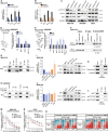
Fig. 4. Inhibition of BCLXL markedly enhances the anti-tumor activity of docetaxel in PDOs and xenograft models.
A–C In vitro activity of BH3-mimetic drugs in combination with docetaxel in PDOs. Left panel: PDOs #2–#6 in Fig. 2H were treated with vehicle, 500 nM docetaxel, BH3-mimetic drug, or both. The concentrations of BCLXLi, MCL1i and BCL2i: PDO #2: 20 nM, PDO #3: 10 nM, PDO #4: 100 nM, PDO #5: 10 nM, PDO #6: 100 nM. Cell viability was determined 72 h later using CellTitre-Glo 3D assays and the mean IC50s ± SD of 3 independent experiments are shown. Right panel: Synergistic activity of different combinations. The expected versus observed effects of BH3-mimetic drugs in combination with docetaxel were calculated using the BLISS model. Unpaired Student’s t-test was used to determine statistical significance. D Schedule and doses of drug treatment for in vivo studies. E Inhibition of BCLXL markedly enhanced the anti-tumor activity of docetaxel in vivo. NCI-N87 xenografts were treated as indicated and tumor growth was summarized. Data shown represent the mean tumor volumes ± SD of 5–6 mice in each group. Two-way ANOVA was used for statistical significance. F Images (top) and quantification of tumor weights (bottom) in the NCI-N87 xenografts. Unpaired Student’s t-test was used for statistical significance. GJ Similar experiments to those in (E, F) were performed in two PDX xenografts.
Fig. 5
Fig. 5. HER2-targeting drugs suppress MCL1 transcription and synergize with BCLXL inhibitor to kill HER2-amplified GC cells.
A–C Effect of HER2-targeting drugs on MCL1 protein levels. NCI-N87 (HER2-amplifed), SNU-216 (HER2-amplified) and GCIY (HER2-nonamplified) were treated with trastuzumab (100 ng/ml) or lapatinib (200 nM) for the indicated time periods. The protein levels of MCL1 were determined by Western blotting. D Blocking ubiquitin- or lysosome-mediated protein degradation failed to reverse the decrease in MCL1 protein levels. NCI-N87 cells were treated with trastuzumab (100 ng/ml), lapatinib (200 nM) alone or together with inhibitors of proteasome (MG132, 5 μM) or lysosome (chloroquine, CQ, 5 μM) for 72 h or 48 h respectively. The protein levels of MCL1 were determined by Western blotting. E, F Effect of trastuzumab and lapatinib on MCL1 mRNA levels. NCI-N87, SNU-216 and GCIY were treated with trastuzumab (100 ng/ml) or lapatinib (200 nM) for the indicated time periods. The mRNA levels of MCL1 were determined by RT-qPCR. G In vitro activity of combining BCLXLi and trastuzumab. The cell viability of NCI-N87, SNU-216 or GCIY 72 h after treatment with 100 ng/ml trastuzumab alone or in combination with indicated concentrations of BCLXL inhibitor was determined. H In vitro activity of combining BCLXLi and lapatinib. The cell viability of NCI-N87, SNU-216, or GCIY 48 h after treatment with lapatinib alone (0–100 nM) or in combination with BCLXL inhibitor was determined. The concentrations of BCLXLi used: 10 nM, 20 nM, and 1 µM for NCI-N87, SNU-216, and GCIY, respectively. I Inhibition of BCLXL markedly enhanced the anti-tumor activity of HER2-targeting drugs in vivo. NCI-N87 xenografts were treated as indicated and tumor growth was summarized. Data shown represents the mean tumor volumes ± SD of 5–6 mice in each group. Two-way ANOVA was used to determine statistical significance. J Images (right) and quantification of tumor weights (left) in the NCI-N87 xenografts. Unpaired Student’s t-test was used to determine statistical significance. Cell viability was determined using the CellTiter-Glo assay; blots in (A–D) are representatives of 2 independent experiments; data in (EH) represent the means ± SD of ≥3 independent experiments.
Fig. 6
Fig. 6. STAT3 and SRF cooperate to mediate the transcriptional response of MCL1 exerted by HER2-targeting drug treatment.
A, B Role of ELK1, STAT3 and SRF in regulating the transcriptional activity of MCL1 under basal conditions and in response to HER2-targeting drugs. SNU-216 cells expressing empty vector or the indicated transcriptional factors were left untreated or treated with 100 ng/ml trastuzumab (A) or 200 nM lapatinib (B) for 72 h or 48 h, respectively. The levels of MCL1 mRNA were determined using RT-qPCR. C Effect of HER2-targeting drugs on STAT3 and SRF expression. SNU-216, NCI-N87 and GCIY were treated with 100 ng/ml trastuzumab or 200 nM lapatinib for 72 h or 48 h, respectively. The protein levels of p-STAT3, total STAT3, and SRF were determined by Western blotting. D, E Dual luciferase reporter assays to identify the binding site(s) of STAT3 and SRF in the MCL1 promoter. Left: diagram depicting the putative binding sites of STAT3 (D) or SRF (E) in the MCL1 promoter predicted by the JASPAR database. Right: Dual luciferase reporter assays conducted in HEK293T. F Reduced binding of STATS and SRF in the MCL1 promoter upon HER2-targeting drug treatments. SNU-216 cells were left untreated or treated with 100 ng/ml trastuzumab or 200 nM lapatinib for 72 h or 48 h, respectively. ChIP was conducted using specific antibodies against STAT3, SRF, and an IgG isotype control. G Co-IP analysis of the interaction of endogenous STAT3 and SRF in SNU-216 cells. H Direct interaction of exogenous STAT3 and SRF. HEK293T cells were transiently transfected with HA-tagged STAT3, Flag-tagged SRF, either individually or both. Equivalent lysates were immunoprecipitated with anti-HA or anti-Flag antibody and immunoblotted with the indicated antibodies. I Effect of overexpressing or deleting STAT3 on SRF expression. The mRNA (left) and protein (right) levels of SRF in SNU-216 cells with STAT3 overexpression or depletion were determined. J Similar experiments to those in panel I were conducted to detect the effect of manipulating SRF on STAT3 expression in SNU-216 cells. K Reduced binding of STAT3 in the SRF promoter upon HER2-targeting drug treatments. Similar experiments to those in (F) were conducted. L Depletion of STAT3 attenuated the binding of SRF in the MCL1 promoter. Similar experiments to those in (F) were performed in SNU-216 cells engineered to express sgSTAT3. ChIP was conducted at 72 h after addition of DOX to induce the deletion of STAT3. 2 sgRNAs were tested. M, N Deleting STAT3 enhanced the sensitivity of GC lines to BCLXL inhibition. SNU-216 (M) and GCIY (N) cells inducibly expressing sgSTAT3 or the sgRNA empty vector were treated with DOX alone, or in combination with indicated concentrations of BCLXLi. Cell viability was determined 48 h later. Two-way ANOVA was used for statistical significance. O STAT3 inhibitor synergized with BCLXLi in GC cell lines, regardless of their p-STAT3 levels. GC cell lines with high p-STAT3 levels (HGC-27, MKN45 and AGS) and the ones with low p-STAT3 levels (SNU-216, NCI-N87, GCIY) were treated with indicated concentrations of BCLXLi and STAT3 inhibitor and cell viability was determined 48 h later. Data in (A), (B), (D), (E), (I, left), (J, left), (MO) represent the means ± SD of ≥3 independent experiments; data in panel (C), (FH), (I, right), (J, right), (K) and (L) are representatives of 2 independent experiments.

References

    1. Bray F, Laversanne M, Sung H, Ferlay J, Siegel RL, Soerjomataram I, et al. Global cancer statistics 2022: GLOBOCAN estimates of incidence and mortality worldwide for 36 cancers in 185 countries. CA Cancer J Clin. 2024;74:229–63. - PubMed
    1. Taieb J, Bennouna J, Penault-Llorca F, Basile D, Samalin E, Zaanan A. Treatment of gastric adenocarcinoma: a rapidly evolving landscape. Eur J Cancer. 2023;195:113370. - PubMed
    1. Lu J, Zheng ZF, Wang W, Xie JW, Wang JB, Lin JX, et al. A novel TNM staging system for gastric cancer based on the metro-ticket paradigm: a comparative study with the AJCC-TNM staging system. Gastric Cancer. 2019;22:759–68. - PubMed
    1. Montero J, Haq R. Adapted to survive: targeting cancer cells with BH3 mimetics. Cancer Discov. 2022;12:1217–32. - PMC - PubMed
    1. Souers AJ, Leverson JD, Boghaert ER, Ackler SL, Catron ND, Chen J, et al. ABT-199, a potent and selective BCL-2 inhibitor, achieves antitumor activity while sparing platelets. Nat Med. 2013;19:202–8. - PubMed

MeSH terms