. 2025 Mar 12;23(1):205.
doi: 10.1186/s12951-025-03275-4.

Injectable hydrogels with ROS-triggered drug release enable the co-delivery of antibacterial agent and anti-inflammatory nanoparticle for periodontitis treatment

Affiliations

Injectable hydrogels with ROS-triggered drug release enable the co-delivery of antibacterial agent and anti-inflammatory nanoparticle for periodontitis treatment

Yujing Zhu et al. J Nanobiotechnology. .

Abstract

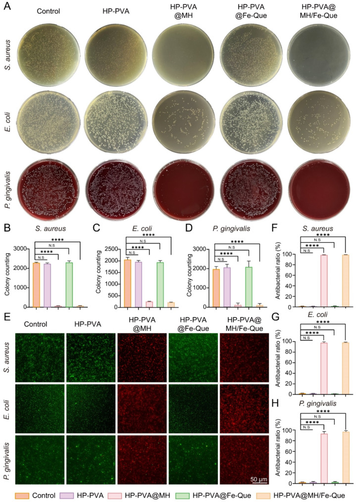
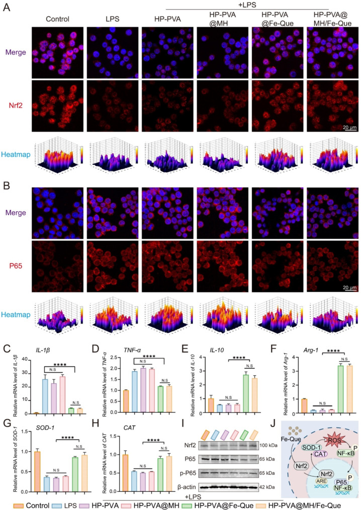
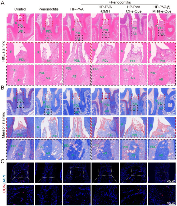
Periodontitis, a chronic inflammatory disease caused by bacteria, is characterized by localized reactive oxygen species (ROS) accumulation, leading to an inflammatory response, which in turn leads to the destruction of periodontal supporting tissues. Therefore, antibacterial, scavenging ROS, reducing the inflammatory response, regulating periodontal microenvironment, and alleviating alveolar bone resorption are effective methods to treat periodontitis. In this study, we developed a ROS-responsive injectable hydrogel by modifying hyaluronic acid with 3-amino phenylboronic acid (PBA) and reacting it with poly(vinyl alcohol) (PVA) to form a borate bond. In addition, the ROS-responsive hydrogel encapsulated the antibacterial agent minocycline hydrochloride (MH) and Fe-Quercetin anti-inflammatory nanoparticles (Fe-Que NPs) for on-demand drug release in response to the periodontitis microenvironment. This hydrogel (HP-PVA@MH/Fe-Que) exhibited highly effective antibacterial properties. Moreover, by modulating the Nrf2/NF-κB pathway, it effectively eliminated ROS and promoted macrophage polarization to the M2 phenotype, reducing inflammation and enhancing the osteogenic differentiation potential of human periodontal ligament stem cells (hPDLSCs) in the periodontal microenvironment. Animal studies showed that HP-PVA@MH/Fe-Que significantly reduced alveolar bone loss and enhanced osteogenic factor expression by killing bacteria and inhibiting inflammation. Thus, HP-PVA@MH/Fe-Que hydrogel had efficient antibacterial, ROS-scavenging, anti-inflammatory, and alveolar bone resorption-alleviation abilities, showing excellent application potential for periodontitis healing.

Keywords: Anti-inflammation; Antibacterial; Drug delivery; Periodontitis; ROS-responsive hydrogels.


Conflict of interest statement

Declarations. Ethics approval and consent to participate: All animal experiments were approved by the animal ethics committee of Southwest Medical University (Document No. 20230705-005). Consent for publication: All authors agree for publication. Competing interests: The authors declare no competing interests.

Figures

Scheme 1
Scheme 1
Schematic diagram of the HP-PVA@MH/Fe-Que hydrogel synthesis and treatment of periodontitis. (A) Synthetic process of Fe-Que NPs. (B) Preparation of HP-PVA@MH/Fe-Que hydrogel. (C) ROS-triggered on-demand release of therapeutic agents in HP-PVA@MH/Fe-Que hydrogel. (D) HP-PVA@MH/Fe-Que hydrogel has multifunctional properties, including antibacterial, regulating Nrf2/NF-κB pathway to inhibit inflammatory and bone absorption relief for treating periodontitis
Fig. 1
Fig. 1
Synthesis and characterization of Fe-Que NPs and HP. (A) Schematic diagram of the synthesis of Fe-Que NPs. (B) Picture of the synthetic materials, including Fe-PVP, Que, and Fe-Que NPs. (C) TEM and (D) Grain size of Fe-Que NPs. (E) UV-vis absorption spectra of FeCl3·6H2O, Que, and Fe-Que NPs. (F) FTIR spectra of Que and Fe-Que NPs. (G) Full XPS spectrum of Fe-Que NPs. (H) High-resolution spectra of ferric ions in Fe-Que NPs. (I) UV-vis absorption spectra and (J) FTIR spectra of HA, PBA, and HP. (K) 1H NMR spectra of HA and HP
Fig. 2
Fig. 2
Preparation and characterization of the HP-PVA@MH/Fe-Que hydrogels. (A) Schematic diagram of the synthesis of hydrogels. (B) Photographs of the HP-PVA@MH/Fe-Que hydrogel before (left) and after (right) gelation. (C) SEM images of the different hydrogels. (D) EDS mapping of the HP-PVA@MH/Fe-Que hydrogel. Photographs of the (E) injectability, (F) shape-adaptability, and (G) adhesion of the HP-PVA@MH/Fe-Que hydrogel. (H) Photographs of the self-healing properties of the HP-PVA hydrogel
Fig. 3
Fig. 3
In vitro antibacterial activity of the HP-PVA@MH/Fe-Que hydrogels. (A) Representative images of S. aureus, E. coli, and P. gingivalis colony-forming units. The pertinent statistical data for the quantification of bacterial colonies of (B) S. aureus, (C) E. coli, and (D) P. gingivalis. (E) The live/dead staining images of S. aureus, E. coli and P. gingivalis. Antibacterial rate of (F) S. aureus, (G) E. coli and (H) P. gingivalis
Fig. 4
Fig. 4
Biocompatibility of the HP-PVA@MH/Fe-Que hydrogels. The Live/Dead staining images of (A) hPDLSCs and (C) RAW264.7 cells for 1, 3, and 5 days, respectively. The CCK-8 assays of (B) hPDLSCs and (D) RAW264.7 cells for 1, 3, and 5 days. (E) Hemolysis ratio of erythrocyte after incubating with the hydrogels
Fig. 5
Fig. 5
Free radical scavenging ability and antioxidant capacity of the HP-PVA@MH/Fe-Que hydrogels under oxidative stress environment. (A) Schematic diagram showing the DPPH free radical scavenging assay. (B) Absorbance, photograph, and (C) DPPH scavenging ratio of the different hydrogels. (D) Schematic diagram showing the ABTS free radical scavenging assay. (E) Absorbance, photograph, and (F) ABTS scavenging ratio of the different hydrogels. (G) DCFH-DA staining, (H) fluorescent quantitative results, (I) Live/dead staining, and (J) CCK-8 assay of the RAW264.7 cell
Fig. 6
Fig. 6
HP-PVA@MH/Fe-Que hydrogels inhibit the polarization of macrophages toward the M1 phenotype while facilitating their polarization toward the M2 phenotype. (A) Schematic diagram showing the effect of the HP-PVA@MH/Fe-Que hydrogels on macrophage polarization. (B) Microscopic view of macrophages. IF images of (C) M1 (iNOS) and (E) M2 (CD206) macrophages maker. Flow cytometry analysis of (D) M1 (CD86) and (F) M2 (CD206) macrophage markers
Fig. 7
Fig. 7
HP-PVA@MH/Fe-Que hydrogels regulate the Nrf2/NF-κB pathway to exert antioxidant and anti-inflammatory effects in vitro. IF images of RAW264.7 cells for (A) Nrf2 and (B) NF-κB. QRT-PCR analysis of the relative mRNA level of (C, D) pro-inflammatory genes IL-1β, TNF-α; (E, F) anti-inflammatory genes IL-10, Arg-1; (G, H) antioxidant enzyme genes SOD-1, CAT. (I) Western blot detection of Nrf2, P65, and p-P65, with β-actin as the internal control. (J) The antioxidant and anti-inflammatory mechanism of HP-PVA@MH/Fe-Que hydrogel was presented as a schematic diagram
Fig. 8
Fig. 8
HP-PVA@MH/Fe-Que hydrogels protected hPDLSCs from ROS attack and promoted osteogenic differentiation in the oxidative microenvironment. (A) Schematic diagram showing the effect of the HP-PVA@MH/Fe-Que on the hPDLSCs’ osteogenic differentiation potential under oxidative stress conditions. (B) Cell morphology of hPDLSCs under oxidative stress condition. (C) ALP staining on day 7 and ARS staining on day 21 of hPDLSCs. (D) IF staining images of Runx2 and OCN in hPDLSCs
Fig. 9
Fig. 9
HP-PVA@MH/Fe-Que hydrogels reduced alveolar bone loss in rat models of periodontitis. (A) Schematic illustration of the treatment procedure for the in vivo study. (B) 3D reconstruction, sagittal images of the alveolar bone in 2 weeks (upper dashed line: CEJ, lower dashed line: ABC, red line: bone loss). (C) Images in 3D reconstruction and 2D sagittal view taken four weeks post-implantation. (D) Assessment of the height of lateral buccal bone loss in quantitative terms and (E) sagittal bone loss of 2 weeks. (F) Quantitative evaluation lateral buccal bone loss height and (G) sagittal bone loss of 4 weeks
Fig. 10
Fig. 10
HP-PVA@MH/Fe-Que hydrogels alleviated the imbalance of periodontal bone homeostasis in rat models of periodontitis after 4 weeks of treatment. (A) Images of H&E staining. (B) Masson’s trichrome staining images of the alveolar bone. (C) IF staining of osteoinductive marker OCN in the alveolar bone. R: root, PDL: periodontal ligament, AB: alveolar bone
Fig. 11
Fig. 11
HP-PVA@MH/Fe-Que hydrogels relieved periodontal tissue inflammation in rat models of periodontitis. (A) Representative IHC staining images of periodontal tissues, including IL-1β and TNF-α. (B) Immunofluorescence staining images of iNOS and CD206 in the area of the alveolar septal region. (C) Immunofluorescence staining images of Nrf2 and P65 in the region of the alveolar septum. (D-I) The corresponding quantitative analysis of IL-1β, TNF-α, iNOS, CD206, Nrf2, and P65

References

    1. Armitage GC. Periodontal diagnoses and classification of periodontal diseases. Periodontol 2000. 2004;34:9–21. 10.1046/j.0906-6713.2002.003421.x. - PubMed
    1. Williams DW, Greenwell-Wild T, Brenchley L, Dutzan N, Overmiller A, Sawaya AP, Webb S, Martin D, Genomics NN, Computational Biology C, et al. Human oral mucosa cell atlas reveals a stromal-neutrophil axis regulating tissue immunity. Cell. 2021;184:4090–e41044015. 10.1016/j.cell.2021.05.013. - PMC - PubMed
    1. Kinane DF, Stathopoulou PG, Papapanou PN. Periodontal diseases. Nat Rev Dis Primers. 2017;3:17038. 10.1038/nrdp.2017.38. - PubMed
    1. Tonetti MS, Chapple ILC. Biological approaches to the development of novel periodontal therapies– Consensus of the seventh European workshop on periodontology. J Clin Periodontol. 2011;38:114–8. 10.1111/j.1600-051X.2010.01675.x. - PubMed
    1. Hathaway-Schrader JD, Novince CM. Maintaining homeostatic control of periodontal bone tissue. Periodontol. 2000. 2021;86:157–187. 10.1111/prd.12368 - PMC - PubMed

MeSH terms