Review
. 2025 Mar 4;17(5):879.
doi: 10.3390/cancers17050879.

The Complexity of Malignant Glioma Treatment

Affiliations
Review

The Complexity of Malignant Glioma Treatment

Linde F C Kampers et al. Cancers (Basel). .

Abstract

Malignant glioma is a highly aggressive, therapeutically non-responsive, and deadly disease with a unique tumor microenvironment (TME). Of the 14 currently recognized and described cancer hallmarks, five are especially implicated in malignant glioma and targetable with repurposed drugs: cancer stem-like cells, in general, and glioma stem-like cells in particular (GSCs), vascularization and hypoxia, metabolic reprogramming, tumor-promoting inflammation and sustained proliferative signaling. Each hallmark drives malignant glioma development, both individually and through interactions with other hallmarks, in which the TME plays a critical role. To combat the aggressive malignant glioma spatio-temporal heterogeneity driven by TME interactions, and to overcome its therapeutic challenges, a combined treatment strategy including anticancer therapies, repurposed drugs and multimodal immunotherapy should be the aim for future treatment approaches.

Keywords: immunotherapy; malignant glioma; tumor microenvironment.


Conflict of interest statement

The authors declare no conflicts of interest.

Figures

Figure 1
Figure 1
Top: An overview of the main contributing factors to the 14 hallmarks of cancer [1,2,3]. In green, the first hallmarks described. In yellow, hallmarks recognized and added in 2011. In blue, four additional hallmarks added in 2022. Legends on the lower left and right side indicate cell types, cytokines, chemokines, growth factors, and other factors vital in tumor development. Dark brown cells of irregular shapes represent tumor cells. Each hallmark has been given an icon, indicated by the numbered images in circles at the end of each hallmark pie slice. These icons will be used to indicate each hallmark in further figures.
Figure 2
Figure 2
A simplified overview of the effect of glioma stem cells. GSCs can self-renew, differentiate and dedifferentiate to different GBM cell types (red arrows). This can be caused by RT or CTx (TMZ) treatment (red bold arrows). Antidepressants can inhibit GSC plasticity. GSCs rely on a permissive TME (black arrows).
Figure 3
Figure 3
A simplified overview of the effects of hypoxia in GBM. GBM Effects are displayed with red arrows, treatment options are shown in bright green. Hypoxia normally leads to autophagy (black arrow) which inhibits the effects of RT and CTx such as TMZ. Chloroquine treatment can inhibit autophagy. Bevacizumab inhibits angiogenesis factor VEGF. Hypoxia also includes upregulation of HIF-1α, which inhibits both CTx effects and the OXPHOS pathway, driving cellular metabolism toward anaerobic fermentation. Mebendazole and melatonin can be used to normalize HIF-1α expression levels.
Figure 4
Figure 4
A simplified overview of metabolic reprogramming in GBM. Normal pathway steps are depicted with black arrows, GBM pathway steps are shown in red. Possible treatment options are depicted in bright green. Glucose is processed through glycolysis to intermediates, ATP and NADH. In cancer cells, an increase in pyruvate dehydrogenase kinase (PDK) inhibits aerobic respiration, favoring an anaerobic fermentation pathway instead. Lactic acid production leads to the acidification of the TME. Treatment with lipoic acid inhibits PDK. Metformin can then be used to inhibit the OXPHOS pathway in all fast-growing cells, specifically targeting tumor cells. Combining TMZ with Metformin treatment can revert chemoresistance.
Figure 5
Figure 5
A simplified overview of the effects of immune response inflammation in GBM. Tumor-specific occurrences are indicated with red arrows, possible treatment options and effects in bright green. Downregulation (downward black arrows) of cell types or cytokines is highlighted in red, and upregulation (upward black arrows) is highlighted in orange. Due to TME acidification, M2 Macrophages and GAMs are upregulated, which inhibits cytotoxic T cell response but increases inflammation-regulating cytokines, developing into an immunosuppressive TME. Especially the upregulation of TGF-β, by both tumor-associated macrophages as well as TCA cycle mutations, further inhibits the innate and adaptive immune system. TCA cycle mutations can be halted with ONC201 treatment, isoselective inhibitors and peptide vaccines. GAMs, especially when derived from bone marrow, also aid angiogenesis. PDL1+ M2 Macrophages target CD8+ T cells via the apoptotic TRAIL pathway, both during CD8+ T cell priming and effector function. Using anti-PDL1 checkpoint inhibitors, this PD1-independent immunosuppression can be inhibited.
Figure 6
Figure 6
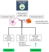
A simplified overview of the effects of the neuron-glioma interactions. The interactions between presynaptic neurons and postsynaptic gliomas drive tumor development through AMPA postsynaptic currents, paracrine signaling factors, or GABAergic synaptic communication. The first can be inhibited with perampanel and the last one with levetiracetam.
Figure 7
Figure 7
Single-cell RNA and protein analysis of myeloid cells in the pontine region of syngeneic allograft mouse models for DMG, indicating PDL1 expression in GAMs compared to healthy cells. Color scales indicate expression levels. Red circles indicate the different tumor–associated grouped cells.
Figure 8
Figure 8
Example of temporal occurrence of individual tumor hallmarks during tumor development, from stage I, Tumorigenesis to stage IV, Invasion. Included is a temporal tumor treatment plan, to combat specific tumor hallmarks throughout malignancy development.
Figure 9
Figure 9
Overview of possible combination strategy trajectory, separated into three treatment phases that allow for adaptation and optimal patient-individualized treatment, based on tumor status and activity. GBM leads to symptoms (indicated as a stethoscope) and suppresses the immune system. Three subsequent treatment phases are proposed to combat the initial tumor and its escape mechanisms, by temporal targeting both, the tumor directly, and its TME. The gray–red–yellow–green background colors reflect treatment effectivity as increased tumor control, also shown as tumor dedifferentiation and size decrease at the top of the figure. Yellow tumor cells represent glioma stem cells, tumor cells in variations of brown (light to dark) represent tumor cells of a different subtype, indicating here how a tumor of a different variation might grow after initial treatment of the main tumor subtype present, and how treatment might push further tumor differentiation.

References

    1. Hanahan D., Weinberg R.A. The Hallmarks of Cancer. Cell. 2000;100:57–70. doi: 10.1016/S0092-8674(00)81683-9. - DOI - PubMed
    1. Hanahan D., Weinberg R.A. Hallmarks of Cancer: The Next Generation. Cell. 2011;144:646–674. doi: 10.1016/j.cell.2011.02.013. - DOI - PubMed
    1. Hanahan D. Hallmarks of Cancer: New Dimensions. Cancer Discov. 2022;12:31–46. doi: 10.1158/2159-8290.CD-21-1059. - DOI - PubMed
    1. Louis D.N., Perry A., Wesseling P., Brat D.J., Cree I.A., Figarella-Branger D., Hawkins C., Ng H.K., Pfister S.M., Reifenberger G., et al. The 2021 WHO Classification of Tumors of the Central Nervous System: A summary. Neuro-Oncology. 2021;23:1231–1251. doi: 10.1093/neuonc/noab106. - DOI - PMC - PubMed
    1. Rouse C., Gittleman H., Ostrom Q.T., Kruchko C., Barnholtz-Sloan J.S. Years of potential life lost for brain and CNS tumors relative to other cancers in adults in the United States, 2010. Neuro-Oncology. 2016;18:70–77. doi: 10.1093/neuonc/nov249. - DOI - PMC - PubMed

LinkOut - more resources