Review
. 2025 Mar 5;17(5):897.
doi: 10.3390/cancers17050897.

Human Papillomavirus-Related Cutaneous Squamous Cell Carcinoma

Affiliations
Review

Human Papillomavirus-Related Cutaneous Squamous Cell Carcinoma

Alejandra Sandoval-Clavijo et al. Cancers (Basel). .

Abstract

The human papillomavirus (HPV) has been associated with the carcinogenesis of cutaneous squamous cell carcinoma (cSCC), especially in immunosuppressed patients. This article reviews the microbiology of HPV and its role in tissue tropism, invasion, and oncogenesis. It also describes possible HPV oncogenic ability due to the inactivation of the host p53 and retinoblastoma protein (pRb) by HPV oncoproteins E6 and E7, producing a suppression of cell cycle checkpoints and uncontrolled cell proliferation that may eventually result in invasive carcinoma. We will focus on β-HPV types and their role in epidermodysplasia verruciformis (EV), as well as α types and their ability to cause cutaneous and mucosal pathology. We also intend to examine the clinical characteristics of cSCC related to HPV and host immunosuppression conditions such as solid organ transplant in order to provide management guidelines for patients with cSCC associated with HPV based on available data. Other topics addressed in this article include particular locations of cSCC, such as nails; the prognosis; the recurrence; therapeutic modalities; and the role of HPV vaccines.

Keywords: Bowen’s disease; cutaneous squamous cell carcinoma; epidermodysplasia verruciformis; human papillomavirus; keratoacanthoma; squamous cell carcinoma.


Conflict of interest statement

The authors declare no conflicts of interest.

Figures

Figure 1
Figure 1
Comparative genome arrangement of α and β genera. Despite having a similar genetic structure, the size and position of the major ORFs can vary; also, β HPV lacks E5 ORF.
Figure 2
Figure 2
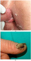
Clinical appearance of HPV-associated SCC. (a) Plaque HPV 16+ in the perianal region, corresponding to in situ SCC. (b) An infiltrative, verrucous plaque in the periungual region with nail plate destruction.
Figure 3
Figure 3
Clinical appearance of different tumors in patients with EV phenotypes. (a) Rounded hyperkeratotic cSCC, with well-defined borders, in the leg of a renal transplant patient with an EV phenotype. (b) Multiple verruco-keratous lesions in a renal transplant patient undergoing treatment with AZA with an EV phenotype. (c) Flat warts in a lung transplant patient with acquired EV. (d) Seborrheic keratosis with a hyperkeratotic verrucous surface. (e) Histopathology of flat warts in a lung transplant patient with acquired EV showing epidermal hyperkeratosis, orthokeratosis, mild papillomatosis, acanthosis, and viral inclusion bodies (scale bar = 100 µm H&E, 10×).
Figure 3
Figure 3
Clinical appearance of different tumors in patients with EV phenotypes. (a) Rounded hyperkeratotic cSCC, with well-defined borders, in the leg of a renal transplant patient with an EV phenotype. (b) Multiple verruco-keratous lesions in a renal transplant patient undergoing treatment with AZA with an EV phenotype. (c) Flat warts in a lung transplant patient with acquired EV. (d) Seborrheic keratosis with a hyperkeratotic verrucous surface. (e) Histopathology of flat warts in a lung transplant patient with acquired EV showing epidermal hyperkeratosis, orthokeratosis, mild papillomatosis, acanthosis, and viral inclusion bodies (scale bar = 100 µm H&E, 10×).
Figure 4
Figure 4
Graphical representation of types of HPV genera associated with mucosal and cutaneous lesions.

References

    1. Que S.K.T., Zwald F.O., Schmults C.D. Cutaneous Squamous Cell Carcinoma: Incidence, Risk Factors, Diagnosis, and Staging. J. Am. Acad. Dermatol. 2018;78:237–247. doi: 10.1016/j.jaad.2017.08.059. - DOI - PubMed
    1. Cerqueira C., Schiller J.T. Papillomavirus Assembly: An Overview and Perspectives. Virus Res. 2017;231:103–107. doi: 10.1016/j.virusres.2016.11.010. - DOI - PMC - PubMed
    1. Nelson C.W., Mirabello L. Human Papillomavirus Genomics: Understanding Carcinogenicity. Tumour Virus Res. 2023;15:200258. doi: 10.1016/j.tvr.2023.200258. - DOI - PMC - PubMed
    1. Van Doorslaer K., McBride A.A. Molecular Archeological Evidence in Support of the Repeated Loss of a Papillomavirus Gene. Sci. Rep. 2016;6:33028. doi: 10.1038/srep33028. - DOI - PMC - PubMed
    1. Gheit T. Mucosal and Cutaneous Human Papillomavirus Infections and Cancer Biology. Front. Oncol. 2019;9:355. doi: 10.3389/fonc.2019.00355. - DOI - PMC - PubMed

LinkOut - more resources