Review
. 2025 Feb 28;15(5):592.
doi: 10.3390/diagnostics15050592.

Unveiling Guyon's Canal: Insights into Clinical Anatomy, Pathology, and Imaging

Affiliations
Review

Unveiling Guyon's Canal: Insights into Clinical Anatomy, Pathology, and Imaging

Sonal Saran et al. Diagnostics (Basel). .

Abstract

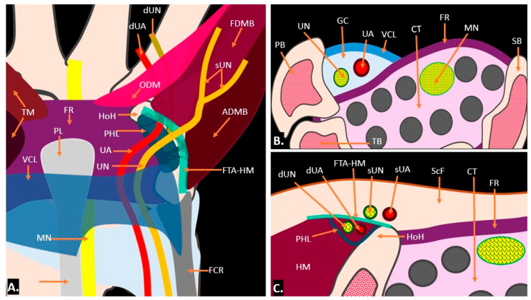
Guyon's canal, or the ulnar tunnel, is a critical anatomical structure at the wrist that houses the ulnar nerve and artery, making it susceptible to various pathological conditions. Pathologies affecting this canal include traumatic injuries, compressive neuropathies like ulnar tunnel syndrome, and space-occupying lesions such as ganglion cysts. Ulnar tunnel syndrome, characterised by numbness, tingling, and weakness in the ulnar nerve distribution, is a prevalent condition that can severely impair hand function. The canal's intricate anatomy is defined by surrounding ligaments and bones, divided into three zones, each containing distinct neural structures. Variations, including aberrant muscles and vascular anomalies, can complicate diagnosis and treatment. Imaging techniques are essential for evaluating these conditions; ultrasound provides real-time, dynamic assessments, while magnetic resonance imaging (MRI) offers detailed visualisation of soft tissues and bony structures, aiding in pre-surgical documentation and pathology evaluation. This review article explores the anatomy, pathologies, and imaging modalities associated with Guyon's canal and underscores the necessity of understanding Guyon's canal's anatomy and associated pathologies to improve diagnostic accuracy and management strategies. By integrating anatomical insights with advanced imaging techniques, clinicians can enhance patient outcomes and preserve hand function, emphasising the need for increased awareness and research in this often-neglected area of hand anatomy.

Keywords: Guyon’s syndrome; MRI scan; diagnostic ultrasound; ganglion cyst; ulnar nerve entrapment at the wrist.


Conflict of interest statement

The authors declare that they have no conflicts of interest.

Figures

Figure 1
Figure 1
Illustration showing the anatomy of Guyon’s canal: (A) presents a frontal view in the coronal plane; (B,C) display axial sections at the proximal and distal levels, respectively. TM = thenar muscle; FR = flexor retinaculum; VCL = volar carpal ligament; MN = median nerve; PL = palmaris longus; UN = ulnar nerve; UA = ulnar artery; FCR = flexor carpi radialis; PHL = pisohamate ligament; HoH = hook of hamate; ODM = opponens digiti minimi; ADMB = abductor digiti minimi brevis; FDMB = flexor digiti minimi brevis; sUN = superficial branches of ulnar nerve; dUN = deep branches of ulnar nerve; dUA = deep branches of ulnar artery; FTA-HM = fibro-tendinous arch of hypothenar muscle; GC = Guyon’s canal; PB = pisiform bone; TB = triquetral bone; SB = scaphoid bone; HM = hypothenar muscles; ScF = subcutaneous fat; FR = flexor retinaculum; and, CT = carpal tunnel.
Figure 2
Figure 2
Illustration depicting the zonal anatomy of Guyon’s canal: Zone 1, located proximally, corresponds to both the motor and sensory components of the ulnar nerve; Zone 2 corresponds to the deep motor branch of the ulnar nerve, which innervates the intrinsic muscles of the hand; and Zone 3 corresponds to the superficial sensory branch, responsible for providing sensation to the ring and little fingers.
Figure 3
Figure 3
Ultrasound anatomy of Guyon’s canal with probe positioning: (A) the Axial Gray-scale Ultrasound image at the level of the proximal Guyon’s canal, at the level of the pisiform bone, containing the ulnar artery (UA) and ulnar nerve (UN), with its floor formed by the flexor retinaculum (open triangles); (B) probe positioning for (A); (C) Axial Gray-scale Ultrasound image at the level of the distal Guyon’s canal, at the level of the hook of the hamate, shows the ulnar nerve dividing into the sensory branch (open triangles) and deep motor branch (open arrow), with the deep motor branch travelling beneath the tendinous arch of the hypothenar muscles; (D) probe positioning for (C); (E) Longitudinal Gray-scale Ultrasound image of the Guyon’s canal shows the ulnar nerve (open arrows) and the underlying flexor retinaculum (an open triangle); and (F) probe positioning for (E).
Figure 4
Figure 4
Axial Gray-scale Ultrasound image at the level of the proximal Guyon’s canal, at the level of the pisiform bone, demonstrates an anatomical variant where an accessory abductor digiti minimi muscle (aADM) overlies the canal. The ulnar nerve (UN) is present within the canal, while the ulnar artery (UA) is positioned volar to the accessory muscle.
Figure 5
Figure 5
Schematic of hypothenar hammer syndrome with ulnar artery aneurysm shown in coronal (A) and axial plane (B).
Figure 6
Figure 6
(A) Axial and (B) longitudinal colour Doppler ultrasound image showing an anechoic cystic lesion within Guyon’s canal, displacing the ulnar artery—likely a simple ganglion cyst.
Figure 7
Figure 7
MRI reveals a well-defined, fluid-filled lesion within Guyon’s canal, consistent with a ganglion cyst (orange arrow), causing compression of the ulnar nerve (UN) (yellow arrow). Image (A) shows an axial view, while image (B) presents a long-axis view.
Figure 8
Figure 8
The image demonstrates ultrasound-guided aspiration of a ganglion cyst, with the needle carefully positioned within the cyst under real-time ultrasound visualisation to ensure precise targeting and safe fluid drainage (A). Graphic overlaying the ultrasound image (B).
Figure 9
Figure 9
(A) T1-weighted coronal, (B) T1-weighted, fat-saturated coronal, and (C) T1-weighted axial and (D) T1-weighted fat-saturated axial MR images showing a typical lipoma (arrow) in Guyon’s canal, appearing as a T1-weighted hyperintense lesion (A,C) filling the Guyon’s canal with complete suppression of fat signals on T1-weighted, fat-saturated sequences (B,D).
Figure 10
Figure 10
(A) T1-weighted coronal and (B) post-contrast T1-weighted, fat-suppressed coronal MR images showing an atypical lipomatous lesion (indicated by orange arrows) in Guyon’s canal, showing T1-weighted hyperintense lesion with hypointense septa in (A) with complete suppression of fat signals and enhancement of the septa in (B).
Figure 11
Figure 11
(A) Gray-scale Axial Ultrasound image showing a well-defined hypoechoic lesion in the region of Guyon’s canal (indicated by callipers). (B) T1-weighted and (C) T2-weighted axial MR images of the same case showing a well-defined lobulated lesion (arrow) appearing iso-to-hypointense to muscle on both (B) T1-weighted and (C) T2-weighted axial MR sequences in keeping with a tenosynovial giant-cell tumour.
Figure 12
Figure 12
(A) T2-weighted, fat-suppressed coronal and (B) T2-weighted, fat-suppressed axial MR images of a neurogenic tumour showing a well-defined elongated lesion (arrow) along the course of the ulnar nerve appearing hyperintense on T2-weighted, fat-suppressed coronal (A) and demonstrating ‘Target-Sign’ with central hypointense signal on axial T2-weighted, fat-suppressed (B) sequences.
Figure 13
Figure 13
(A) Plain radiograph showing a radio-dense lesion located anterior to the pisiform bone. (B,C) Gray-scale Axial Ultrasound images of the same patient showing hyperechoic foci (orange arrow) at the flexor carpi ulnaris insertion over the pisiform bone, in keeping with flexor carpi ulnaris (FCU) calcific tendinopathy. (C) Gray-scale Longitudinal Ultrasound image showing calcium extending into the adjacent soft tissue. Ultrasound image with colour Doppler overlay demonstrates increased vascularity within the flexor carpi ulnaris (FCU) tendon and surrounding soft tissues, suggesting active inflammation.
Figure 14
Figure 14
A case of idiopathic right ulnar neuropathy is illustrated. Image (A) shows an increased cross-sectional area of the right ulnar nerve compared to the left side (B), with a loss of the fascicular pattern. Image (C) presents a long-axis view of both wrists, highlighting the thickened right ulnar nerve in contrast to the normal left ulnar nerve (D). There is no evidence of a clear compressive cause.
Figure 15
Figure 15
(A) T1-weighted axial, (B) T2-weighted, fat-suppressed axial, and (C) T2-weighted, fat-suppressed coronal MR images of an arteriovenous malformation (indicated by the orange arrow) in the region of Guyon’s canal. The lesion appears hypointense on the T1-weighted sequence (A), appears hyperintense on T2-weighted, fat-suppressed sequences, (B) and demonstrates continuity with vessels (blue arrows) (C).
Figure 16
Figure 16
(A) T2-weighted, fat-suppressed coronal and (B) T2-weighted, fat-suppressed axial MR images reveal the tortuous irregular path of the ulnar artery (orange arrow) in the region of Guyon’s canal, resulting in ulnar neuropathy and causing neuropathic symptoms.
Figure 17
Figure 17
(A) Clinical photograph of a 20-year-old male patient presenting with a penetrating injury scar (purple arrow) in the region of Guyon’s canal with possible injury to the left-sided ulnar nerve; (B) Gray-scale Longitudinal Ultrasound image in the region of Guyon’s canal reveals a transected left-sided ulnar nerve with stump neuroma formation (orange arrow); and (C,D) Gray-scale Axial Ultrasound images show denervation atrophy of the left hypothenar muscles (HM) [D] compared to the contralateral normal right-sided muscles (C).
Figure 18
Figure 18
Colour Doppler longitudinal ultrasound image showing ulnar arterial pseudoaneurysm (orange arrow) in a case of hypothenar hammer syndrome.

References

    1. Guyon F.C. Note on a special canal in the anterior annular ligament of the carpus for the passage of the ulnar artery. Comptes Rendus Soc. Biol. 1861;12:92–94.
    1. Ramage J.L., Varacallo M. StatPearls. StatPearls Publishing; Treasure Island, FL, USA: 2020. Anatomy, shoulder and upper limb, hand Guyon canal. - PubMed
    1. Coraci D., Loreti C., Piccinini G., Doneddu P.E., Biscotti S., Padua L. Ulnar neuropathy at wrist: Entrapment at a very “congested” site. Neurol. Sci. 2018;39:1325–1331. doi: 10.1007/s10072-018-3446-7. - DOI - PubMed
    1. Aleksenko D., Varacallo M. StatPearls [Internet] StatPearls Publishing; Treasure Island, FL, USA: 2023. Guyon Canal Syndrome. - PubMed
    1. Becker R.E., Manna B. StatPearls [Internet] StatPearls Publishing; Treasure Island, FL, USA: 2023. Anatomy, Shoulder and Upper Limb, Ulnar Nerve. - PubMed

LinkOut - more resources