

Single-Cell RNA Sequencing Reveals an Atlas of Meihua Pig Testis Cells
- PMID: 40076035
- PMCID: PMC11899385
- DOI: 10.3390/ani15050752
Single-Cell RNA Sequencing Reveals an Atlas of Meihua Pig Testis Cells
Abstract
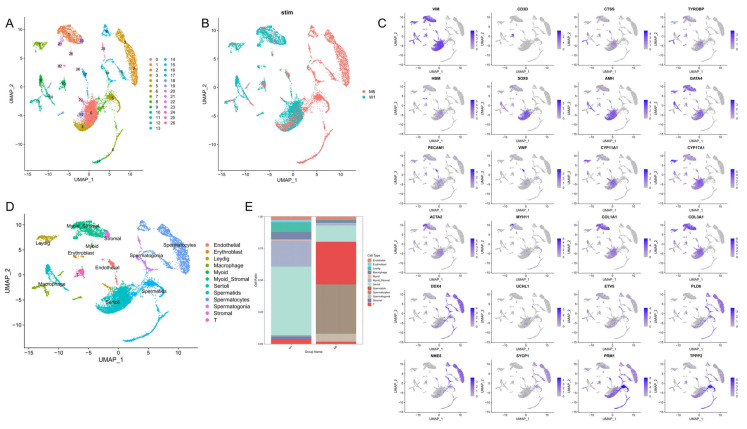
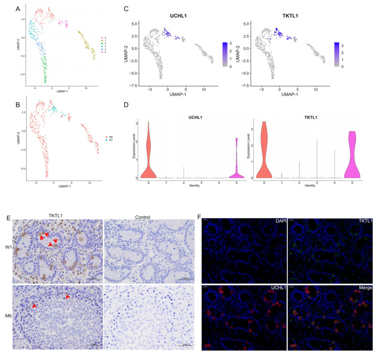
Mammalian spermatogenesis is a complex biological process that is regulated by multiple types of cells. The heterogeneity of these cells poses a challenge for analyzing different cell types at different developmental stages. To characterize the transcriptomic landscape of porcine spermatogenesis and identify potential marker genes for spermatogonia, an unbiased transcriptomic study of spermatogenesis in neonatal and sexually mature six-month-old Meihua pigs was performed using 10× Genomics single-cell RNA sequencing (scRNA-seq). Through the collection of scRNA-seq data from 13,839 cells from Meihua pig testes, three germ cells (spermatogonia, spermatocytes and spermatids) and eight somatic cells (Sertoli cells, Leydig cells, myoid/stromal cells, endothelial cells, T cells/macrophages and erythroblasts) were identified. Pseudo-timing analysis showed that myoid cells and stromal cells originated from common progenitors in Meihua pigs. Functional enrichment analysis revealed that the differentially expressed genes (DEGs) in testicular somatic cells were enriched in the pathways of Ribosome, Oxidative phosphorylation, Protein processing in endoplasmic reticulum, Retrograde endocannabinoid signaling, Cellular senescence and Insulin signaling. Meanwhile, in the three different germ cells, except for pathways which were the same as the first three pathways for somatic cells, DEGs were also enriched in the Spliceosome, Cell cycle, Autophagy and Mitophagy pathways. Furthermore, the candidate marker gene TKTL1 in spermatogonia was identified using immunohistochemistry and immunofluorescence. In conclusion, we collected transcription datasets and constructed single-cell developmental maps of germ cells and somatic cells during the testicular development of Meihua pigs, which provided new insights into the spermatogenesis of Meihua pigs and the development of various types of cells in their testes.
Keywords: Meihua pig; TKTL1; single-cell transcriptome sequencing; testis.
Conflict of interest statement
The authors declare no conflicts of interest.
Figures


References
Grants and funding
- 2021ZDZX4040/Key Fields Special Project for Ordinary Universities in Guangdong Province
- 2022-XJS-00-002/Provincial Rural Revitalization Strategy special fund seed industry revitalization project
- 230403148031043/Shaoguan Science and Technology Plan Project
- 432-9900064501/The Doctoral Initiation Fund of Shaoguan University
- 230324108035353/2023 Shaoguan City Social Development Science and Technology Collaborative Innovation System Construction project
LinkOut - more resources
Full Text Sources
