Review
. 2025 Feb 21;26(5):1851.
doi: 10.3390/ijms26051851.

A Comprehensive Review of Fc Gamma Receptors and Their Role in Systemic Lupus Erythematosus

Affiliations
Review

A Comprehensive Review of Fc Gamma Receptors and Their Role in Systemic Lupus Erythematosus

Jesús Sepúlveda-Delgado et al. Int J Mol Sci. .

Abstract

Receptors for the immunoglobulin G constant fraction (FcγRs) are widely expressed in cells of the immune system. Complement-independent phagocytosis prompted FcγR research to show that the engagement of IgG immune complexes with FcγRs triggers a variety of cell host immune responses, such as phagocytosis, antibody-dependent cell cytotoxicity, and NETosis, among others. However, variants of these receptors have been implicated in the development of and susceptibility to autoimmune diseases such as systemic lupus erythematosus. Currently, the knowledge of FcγR variants is a required field of antibody therapeutics, which includes the engineering of recombinant soluble human Fc gamma receptors, enhancing the inhibitory and blocking the activating FcγRs function, vaccines, and organ transplantation. Importantly, recent interest in FcγRs is the antibody-dependent enhancement (ADE), a mechanism by which the pathogenesis of certain viral infections is enhanced. ADEs may be responsible for the severity of the SARS-CoV-2 infection. Therefore, FcγRs have become a current research topic. Therefore, this review briefly describes some of the historical knowledge about the FcγR type I family in humans, including the structure, affinity, and mechanism of ligand binding, FcγRs in diseases such as systemic lupus erythematosus (SLE), and the potential therapeutic approaches related to these receptors in SLE.

Keywords: Fc gamma receptor; Fc?R; FcgR; FcgRIIIa; FcgRIIIb; FcgRIIa; SLE; autoimmune disease; autoimmunity; phagocytosis.


Conflict of interest statement

The authors declare no conflict of interest.

Figures

Figure 2
Figure 2
Structure of Fc gamma receptors. Fc receptors are composed of an alpha backbone (α), where the activation domain is located; the immunoreceptor tyrosine-based activation motifs (ITAMs), as is the case for FcγRIIa. For FcγRIa and FcγRIIIa, there are accessory chains, such as gamma (γ) and others, which are the carriers of the ITAMs necessary for signaling. The only inhibitory receptor, FcγRIIb, contains immunoreceptor tyrosine-based inhibitory motifs (ITIMs). Each receptor is composed of two immunoglobulin-like domains, with the exception of FcγRIa, which is composed of three domains that favor high-affinity characteristics.
Figure 1
Figure 1
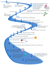
Timeline of Fcγ receptors research. The figure below shows a timeline on the research of Fc gamma receptors. It is a basic and informative line showing the evolution of research on the subject; important points may not have been included in the figure. Dates and references: 1965 [7], 1966 [8], 1967 [9], 1968 [10,11], 1970 [12,13,14], 1972 [15], 1975 [16], 1976 [17], 1977 [18], 1979 [19], 1980 [20], 1982 [21], 1983 [22], 1984 [6], 1988 [23,24], 1989 [25,26,27], 1995 [28], 1996 [29], 1998 [30], 1999 [31], 2000 [32], 2004 [33], 2008 [34], 2013 [35], 2016 [36], 2017 [37], 2019 [38], 2020 [39], 2022 [40], 2023 [41], 2024 [42].
Figure 3
Figure 3
FcγRIIIb receptor polymorphism distribution in exon 3 (EC1): The positions of the nucleotides that have changes can be distinguished according to AJ581669.1, which was generated and used in the initial studies of the receptor. The classification of clinical significance (ClinVar) is also shown, and some cases considered pathogenic to date are under review. The frequency of the variant, according to the database of the 1000 genomes; the changes, according to the single nucleotide variants database; and the name of the variant, according to the global database of all SNPs, are shown. Additionally, the NCBI Reference Sequence Database (RefSeq) is included to identify the location of variants on the chromosome (NC_000001.11), gene (NG_032926.1), protein (NP_001231682.2), and mRNA (AJ581669.1). Modified from [90].
Figure 4
Figure 4
FcγRIIIb signaling pathway (NETosis pathway, red lines and arrows): Due to the lack of ITAMs, the initial steps of signaling are not yet known in detail; however, part of the signaling pathway associated with the formation of NETs has recently been described. The signaling in neutrophils of SLE patients might start from the immune complexes of autoantibodies (the figure represents autoantibody complexes with autoantigens like double-stranded DNA or nuclear proteins, which is common in SLE.) binding to the receptor. Currently, what is known about the pathway has been obtained from in vitro tests. Upon FcγRIIIb IC binding or receptor activation, the Syk and TAK 1 kinases are activated. These enzymes trigger the MEK/ERK signaling cascade. ERK signaling leads to the activation of the NADPH oxidase complex for ROS production, which is required to induce NET formation. PKC is involved in MEK/ERK pathway activation. Also, the nuclear factor Elk-1 gets phosphorylated in the nucleus by a mechanism independent of ERK. The FcγRIIIb activation promotes a pro-adhesive phenotype and enhances NETs; the contribution to phagocytosis is minimal, and phosphorylation of ERK is much more efficient in the nucleus. It favors the expression of beta 2 integrins. FcγRs signaling pathways: FcγRs activating receptors bind to immune complexes, facilitating cross-linking and intracytoplasmic activation, for which tyrosine kinases of the Src family are activated and phosphorylate tyrosine residues in ITAM on the alpha chains of the receptor. Syk, an enzyme with tyrosine kinase activity, is activated by Src. SYK phosphorylates multiple substrates, including SOS a guanine nucleotide exchange factor that activates the Ras-Raf-MEK-ERK (MAPK) pathway, which facilitates the exchange of GDP by GTP in Ras. Ras, a GTPase enzyme, activates Raf, which then phosphorylates and activates MEK, which, in turn, phosphorylates ERK. ERK activates NF-κB. FcγRIIa (Phagocytosis pathway, blue lines and arrows): Additionally, Syk activates PI3K, which produces PIP3 from the phosphorylation of PIP2 in the cell membrane, then PIP3 binds to BTK, a kinase that activates small GTPases, such as Rho and Rac. These GTPases are involved in reorganization of the actin cytoskeleton during phagocytosis. In addition, PIP3 activates PLCγ, which produces the second messengers DAG and IP3. DAG activates PKC. PKC activates the NADPH-oxidase complex to produce ROS. IP3 binds to IP3R in the endoplasmic reticulum to release Ca2+ into the cytoplasm. Finally, this señalización promotes a phagocytic phenotype, cytosol phosphorylation of ERK, oxidative stress, and antibody-dependent cellular cytotoxicity(depending on the cell type). Abbreviations or molecule’s function: A question mark (?) indicates an unknown mechanism of activation. Syk: spleen tyrosine kinase; TAK 1: TGF-beta activated kinase 1; MEK: mitogen-activated protein kinase; ERK: extracellular signal-regulated kinase; PKC: protein kinase C; Sos: son of sevenless (a guanine nucleotide exchange factor); Ras: a GTPase; Raf: a serine/threonine kinase; Elk-1: ETS-like gene 1, a transcription factor. BTK: Bruton’s tyrosine kinase. DAG: diacylglycerol, NF-κB: nuclear factor kappa B; PI3 K: phosphoinositide 3-kinase; PLCγ: phospholipase C gamma; IP3: inositol 1,4,5-trisphosphate. Modified from [126]. Adaptation of figure and text mechanisms [36,126,130,131,132,133].

References

    1. Grundy H.O., Peltz G., Moore K.W., Golbus M.S., Jackson L.G., Lebo R.V. The polymorphic Fc? receptor II gene maps to human chromosome 1q. Immunogenetics. 1989;29:331–335. doi: 10.1007/BF00352843. - DOI - PubMed
    1. Dietzsch E., Osman N., McKenzie I.F.C., Garson M., Hogarth P.M. The human FCG1 gene encoding the high-affinity Fc?RI maps to chromosome 1q21. Immunogenetics. 1993;38:307–309. doi: 10.1007/BF00188811. - DOI - PubMed
    1. Takai S., Kasama M., Yamada K., Kai N., Hirayama N., Namiki H., Taniyama T. Human high-affinity Fc?RI (CD64) gene mapped to chromosome 1q21.2-q21.3 by fluorescence in situ hybridization. Hum. Genet. 1994;93:13–15. doi: 10.1007/BF00218905. - DOI - PubMed
    1. Peltz G.A., Grundy H.O., Lebo R.V., Yssel H., Barsh G.S., Moore K.W. Human Fc gamma RIII: Cloning, expression, and identification of the chromosomal locus of two Fc receptors for IgG. Proc. Natl. Acad. Sci. USA. 1989;86:1013–1017. doi: 10.1073/pnas.86.3.1013. - DOI - PMC - PubMed
    1. de Wit T.P.M., Suijkerbuijk R.F., Capel P.J.A., van Kessel A.G., van de Winkel J.G.J. Assignment of three human high-affinity Fcγ receptor I genes to chromosome 1, band q21.1. Immunogenetics. 1993;38:57–59. doi: 10.1007/BF00216392. - DOI - PubMed

LinkOut - more resources