. 2025 Feb 25;26(5):1994.
doi: 10.3390/ijms26051994.

Impact of Minimally Manipulated Cell Therapy on Immune Responses in Radiation-Induced Skin Wound Healing

Affiliations

Impact of Minimally Manipulated Cell Therapy on Immune Responses in Radiation-Induced Skin Wound Healing

Victoria A Shestakova et al. Int J Mol Sci. .

Abstract

The current treatment of radiation-induced skin wounds utilizes mainly conventional therapies, including topical steroids, creams, ointments, and hydrogel dressings, which do not take into account the immunologic changes that occur in the skin after radiation exposure. Therefore, it is relevant to consider alternative therapies and their impact on changes in the immune landscape of the skin. The aim of this study was to investigate the effect of allogeneic minimally manipulated keratinocytes and fibroblasts on rat skin repair and the development of immune responses. We found that the use of cell therapy compared to treatment with syntazone ointment and no treatment resulted in faster healing and a reduction in the size of radiation-induced skin wounds, area of inflammation, and edema. Additionally, in the group receiving the cell therapy application, there was an observed increase in the number of mast cells (MCs), activation of MC interaction with M2 macrophages, a reduction in the direct contact of MCs with the vascular bed, an increase in the content of collagen fibers due to the intensification of collagen fibrillogenesis, and a restoration of their histotopographic organization. Thus, the positive effect of cell therapy based on allogeneic minimally manipulated keratinocytes and fibroblasts on skin regeneration indicated that it can be used in clinical practice to improve the effectiveness of rehabilitation after radiation therapy.

Keywords: cell therapy; immune responses; ionizing radiation; mast cells; minimally manipulated cells; radiation injury; regenerative medicine; skin.


Conflict of interest statement

The authors declare no conflicts of interest.

Figures
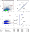
Figure 1
Figure 1
Assessment of viability of cell fractions using flow cytometry by staining with fluorescent dye DAPI: (A) exclusion of cellular debris; (B) exclusion of doublets and highlighting of the area of single cells; (C) live and dead cells; and (D) histogram of the distribution of live and dead cells.
Figure 2
Figure 2
Results of flow cytometry by direct and lateral light scattering: (A) event density plot with selected polygonal regions “All cells”, “Keratinocytes” and “Fibroblasts”; (B) event density plot reflecting the nuclei of populations; (C) histogram of statistical distribution of cells by the number of events attributable to the regions “Keratinocytes” and “Fibroblasts”; and (D) distribution of events by populations.
Figure 3
Figure 3
Skin appearance of rats after irradiation: (A) radial skin wounds development from day 1 to day 9, the area of radial lesion development is highlighted in the circle; and (B) radial skin wounds from day 11 to day 16 during application of Cell therapy, Standard therapy and Negative control.
Figure 4
Figure 4
Hematoxylin and eosin staining: (A) non-irradiated and untreated native rat skin sample (control group); (B) skin sample of rats with radiation-induced skin wounds at the time of wet epidermitis development; (C) skin sample of rats with radiation-induced skin wounds without treatment at the end of the experiment (negative control group); (D) skin sample of rats treated with standard therapy; and (E) skin sample of rats treated with cell therapy. Scale bar 200 μm.
Figure 5
Figure 5
Picro Mallory staining: (A) non-irradiated and untreated native rat skin sample (control group); (B) skin sample of rats with radiation-induced skin wounds at the time of wet epidermitis development; (C) skin sample of rats with radiation-induced skin wounds without treatment at the end of the experiment (negative control group); (D) skin sample of rats treated with standard therapy; and (E) skin sample of rats treated with cell therapy.
Figure 6
Figure 6
A combination of silver impregnation with toluidine blue staining: (A,A’) non-irradiated and untreated native rat skin sample (control group); (B,B’) skin sample of rats with radiation-induced skin wounds at the time of wet epidermitis development; (C) skin sample of rats with radiation-induced skin wounds without treatment at the end of the experiment (negative control group); (D,D’) skin sample of rats treated with standard therapy; and (E,E’) skin sample of rats treated with cell therapy. Scale bar: (AD)—600 μm, (E)—400 μm, others—5 μm.
Figure 7
Figure 7
Histochemical equivalents of targeting mast cells to targets of the tissue microenvironment of the rat dermis of different experimental groups. Methods included (AI) Giemsa staining and (JL) picro Mallory staining protocols: (AD) non-irradiated and untreated native rat skin sample (control group). The secretion of large granules with metachromasia (indicated by arrow) to thin collagen fibers of the papillary layer (A), fibroblast (B), capillary wall (C) and collagen fiber bundles of the reticular layer of the dermis (D) was determined; (E,F) skin sample of rats with radiation-induced skin wounds at the time of wet epidermitis development; (E) intercellular interaction of mast cells and neutrophil (arrow), against the background of accumulation of granular leukocytes in the venous section of the microcirculatory channel with signs of transendothelial migration to the tissue microenvironment of the dermis (white arrow); (F) interaction of mast cells with each other (arrow) and neutrophil granulocyte (white arrow); (G) skin sample of rats with radiation-induced skin wounds without treatment at the end of the experiment (negative control group). Co-localization of mast cell with eosinophil (arrow); (H) skin sample of rats treated with standard therapy; (H’) in a magnified fragment of (H), secretory granules in the extracellular matrix are detected (arrow). Skin sample of rats treated with cell therapy: (I) high mast cell content (indicated by arrow); (J) signs of targeted secretion of mast cell mediators to fibroblast (indicated by arrow) and collagen fiber (indicated by white arrow); and (KL) loci with active collagen fibrillogenesis are identified in the pericellular space of mast cells (arrow). Scale bar: (H,I)—50 μm, others—5 μm.
Figure 8
Figure 8
Expression and target secretion of mast cell tryptase in the skin of rats of different groups. Methods: Combined immunohistochemical and histochemical staining: (A,B) non-irradiated and untreated native rat skin sample (control group): (A) co-localization of mast cells with moderate (arrow), minimal (double arrow) and complete absence (white arrow) of tryptase expression; and (B) localization of type 2 macrophages in the connective tissue base of the skin (arrow); (C,D) skin samples of rats with radiation-induced skin wounds at the time of wet epidermitis development: (C) αSMA+ cells within the vasculature of the skin dermis (arrow); (D) targeted secretion of tryptase to structural components of the venule wall; (E) the skin sample of rats with radiation-induced skin wounds without treatment at the end of the experiment (negative control group), increased tryptase expression in most mast cells (arrow), intercellular interaction (white arrow); (F,G) skin samples of rats treated with standard therapy: (F) close co-localization of a degranulated mast cell with the microcirculatory channel of the skin dermis (arrow); and (G) mast cells are filled with granules with high tryptase content (arrow); (H,I) skin sample of rats treated with cell therapy: (H) most mast cells express tryptase; and (I) mast cell co-localized secretory granules with different levels of tryptase content, including high (arrow), peripheral localization of tryptase is identified in secretory granules. Scale bar: (B,C)—50 μm, others—5 μm.
Figure 9
Figure 9
The content of M2-macrophages and their interaction with mast cells and their secretory granules: (A) monoplex and multiplex immunohistochemical staining; and (B) combined immunohistochemical (CD163 detection) and immunohistochemical (toluidine blue) staining. Arrow indicates mast cell-macrophage type 2 interaction loci. Scale bar: (marked by asterisk (*))—50 μm, others—5 μm.
Figure 10
Figure 10
Collection of skin biomaterial using a biopsy punch: (A) dead rat with depilated skin on its back; (B) extraction of a skin biopsy specimen with a 7.5 mm biopsy punch; (C) appearance of wounds created on the rat’s back with the biopsy punch; and (D) skin biopsy specimen containing epidermal and dermal cells. Created with Biorender.com.

References

    1. Gieringer M., Gosepath J., Naim R. Radiotherapy and wound healing: Principles, management and prospects. Oncol. Rep. 2011;26:299–307. doi: 10.3892/or.2011.1319. - DOI - PubMed
    1. Iddins C.J., DiCarlo A.L., Ervin M.D., Herrera-Reyes E., Goans R.E. Cutaneous and local radiation injuries. J. Radiol. Prot. 2022;2:011001. doi: 10.1088/1361-6498/ac241a. - DOI - PMC - PubMed
    1. Aliper A.M., Bozdaganyan M.E., Sarkisova V.A., Veviorsky A.P., Ozerov I.V., Orekhov P.S., Korzinkin M.B., Moskalev A., Zhavoronkov A., Osipov A.N. Radioprotectors.org: An open database of known and predicted radioprotectors. Aging. 2020;12:15741–15755. doi: 10.18632/aging.103815. - DOI - PMC - PubMed
    1. Christy B.A., Herzig M.C., Wu X., Mohammadipoor A., McDaniel J.S., Bynum J.A. Cell Therapies for Acute Radiation Syndrome. Int. J. Mol. Sci. 2024;25:6973. doi: 10.3390/ijms25136973. - DOI - PMC - PubMed
    1. Yang P., Zhang S., Yan T., Li F., Zhang S. The Therapeutic Application of Stem Cells and Their Derived Exosomes in the Treatment of Radiation-Induced Skin Injury. Radiat. Res. 2023;199:182–201. doi: 10.1667/RADE-22-00023.1. - DOI - PubMed

LinkOut - more resources