

LRRK2 in Drosophila Melanogaster Model: Insights into Cellular Dysfunction and Neuroinflammation in Parkinson's Disease
- PMID: 40076730
- PMCID: PMC11900240
- DOI: 10.3390/ijms26052093
LRRK2 in Drosophila Melanogaster Model: Insights into Cellular Dysfunction and Neuroinflammation in Parkinson's Disease
Abstract
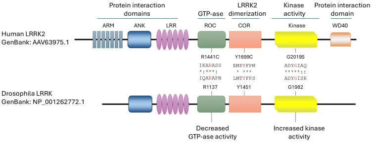
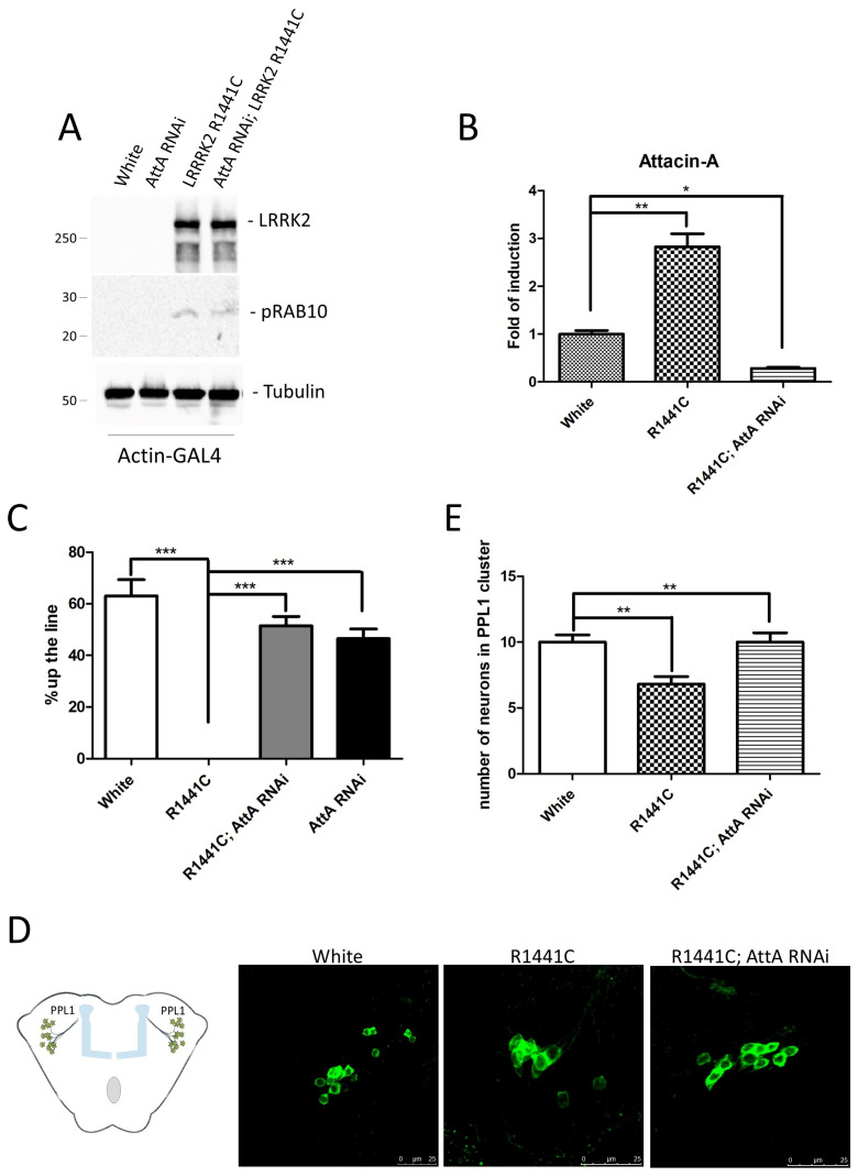
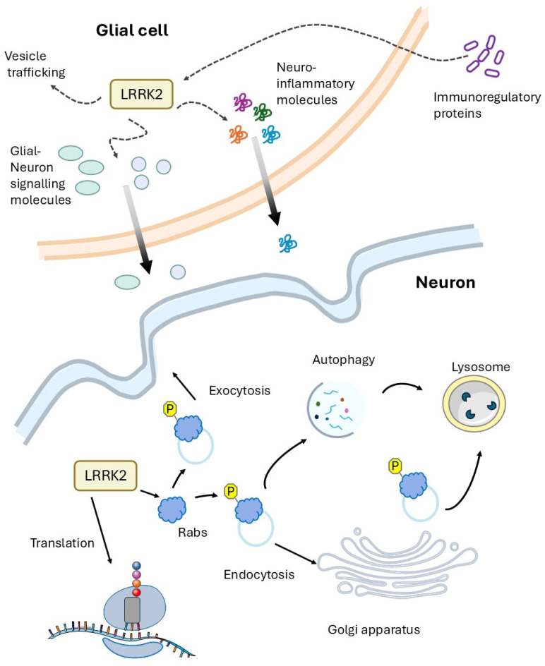
Parkinson's disease (PD) is a fatal neurodegenerative disease for which there are no still effective treatments able to stop or slow down neurodegeneration. To date, pathological mutations in the leucine-rich repeat kinase 2 (LRRK2) gene have been identified as the major genetic cause of PD, although the molecular mechanism responsible for the loss of dopaminergic neurons is still cryptic. In this review, we explore the contribution of Drosophila models to the elucidation of LRRK2 function in different cellular pathways in either neurons or glial cells. Importantly, recent studies have shown that LRRK2 is highly expressed in immunocompetent cells, including astrocytes and microglia in the brain, compared to neuronal expression. LRRK2 mutations are also strongly associated with the development of inflammatory diseases and the production of inflammatory molecules. Using Drosophila models, this paper shows that a genetic reduction of the inflammatory response protects flies from the neurodegeneration induced by LRRK2 pathological mutant expression.
Keywords: Attacin-A; Drosophila; LRRK2; Parkinson’s disease.
Conflict of interest statement
The authors declare no conflicts of interest.
Figures
Similar articles
-
Human R1441C LRRK2 regulates the synaptic vesicle proteome and phosphoproteome in a Drosophila model of Parkinson's disease.Hum Mol Genet. 2016 Dec 15;25(24):5365-5382. doi: 10.1093/hmg/ddw352. Hum Mol Genet. 2016. PMID: 27794539 Free PMC article.
-
The modifying effect of mutant LRRK2 on mutant GBA1-associated Parkinson disease.Hum Mol Genet. 2025 Jul 3;34(14):1184-1203. doi: 10.1093/hmg/ddaf062. Hum Mol Genet. 2025. PMID: 40315377 Free PMC article.
-
LRRK2 regulates ferroptosis through the system Xc-GSH-GPX4 pathway in the neuroinflammatory mechanism of Parkinson's disease.J Cell Physiol. 2024 May;239(5):e31250. doi: 10.1002/jcp.31250. Epub 2024 Mar 13. J Cell Physiol. 2024. PMID: 38477420
-
LRRK2 and mitochondria: Recent advances and current views.Brain Res. 2019 Jan 1;1702:96-104. doi: 10.1016/j.brainres.2018.06.010. Epub 2018 Jun 9. Brain Res. 2019. PMID: 29894679 Free PMC article. Review.
-
Leucine-rich repeat kinase 2-related functions in GLIA: an update of the last years.Biochem Soc Trans. 2021 Jun 30;49(3):1375-1384. doi: 10.1042/BST20201092. Biochem Soc Trans. 2021. PMID: 33960369 Review.
References
-
- Healy D.G., Falchi M., O’Sullivan S.S., Bonifati V., Durr A., Bressman S., Brice A., Aasly J., Zabetian C.P., Goldwurm S., et al. Phenotype, Genotype, and Worldwide Genetic Penetrance of LRRK2-Associated Parkinson’s Disease: A Case-Control Study. Lancet Neurol. 2008;7:583–590. doi: 10.1016/S1474-4422(08)70117-0. - DOI - PMC - PubMed
Publication types
MeSH terms
Substances
Grants and funding
LinkOut - more resources
Full Text Sources
Medical
Molecular Biology Databases
