. 2025 Feb 27;18(5):1065.
doi: 10.3390/ma18051065.

Pure and Doped Brushite Cements Loaded with Piroxicam for Prolonged and Constant Drug Release

Affiliations

Pure and Doped Brushite Cements Loaded with Piroxicam for Prolonged and Constant Drug Release

Marcella Bini et al. Materials (Basel). .

Abstract

The increase in life expectancy has led to a rise of musculoskeletal disorders. Calcium phosphate cements (CPCs), thanks to some amazing features such as the ability to harden in vivo, bioactivity, and resorbability, are promising candidates to treat these diseases, notwithstanding their poor mechanical properties. We aimed to synthesise pure and barium- or silicon-doped brushite-based CPCs loaded with piroxicam to study the effects of the substitution on physical-chemical and pharmaceutical properties before and after cement immersion in phosphate buffer for different time periods. Our results demonstrated that piroxicam became amorphous in the hardened cements. The dopants did not change the brushite structure or its lamellar morphology, while both Ba and Si additions improved the initial Young's modulus compared to the pure cement, and the opposite trend was observed for compressive strength. Both the compressive strength and the elastic modulus decreased for the samples immersed in solution compared to the non-immersed samples, with stabilisation as the number of days increased. After 7 days, the whole drug amount was released, with a slower and constant kinetic for the Ba-doped cements compared to the pure and Si-doped ones.

Keywords: brushite phase; calcium phosphate cements; dissolution rate; doping; mechanical measurements; piroxicam.


Conflict of interest statement

The authors declare no conflicts of interest.

Figures

Scheme 1
Scheme 1
Molecular structure of piroxicam.
Figure 1
Figure 1
XRD patterns of β-TCP, β-TCP-Ba, and β-TCP-Si. The bars of the expected peak positions of β-Ca3(PO4)2 (red) are also reported.
Figure 2
Figure 2
FT-IR spectra of β-TCP, β-TCP-Ba, and β-TCP-Si in the 4000–600 cm−1 spectral range. In the inset, the enlargement of the 1300–700 cm−1 spectral range is shown.
Figure 3
Figure 3
SEM images of β-TCP (A), β-TCP-Ba (B), and β-TCP-Si (C).
Figure 4
Figure 4
XRD patterns of pure and doped cements without (A) and with (B) piroxicam loading. The brushite red bars and the stars marking the main peaks of monetite are also reported.
Figure 5
Figure 5
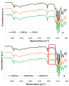
FT-IR spectra of pure and doped cements without drug (A) and loaded with piroxicam (B). The red box highlights the main drug peaks. The spectra were normalised considering the main peak of cement at about 1057 cm−1 as a reference (see text).
Figure 6
Figure 6
SEM images, at different magnifications, of cements without drugs: CEM (A,B), CEM-Ba (C,D), and CEM-Si (E,F). The magnifications are 300× (A,C,E) and 6000× (B,D,F). For the sake of simplicity, the bar of magnification is indicated in the (E,F) images and is valid for images at the same magnification.
Figure 7
Figure 7
SEM images, at different magnifications, of cements with drugs: CEM-Prx (A,B), CEM-Ba-Prx (C,D), and CEM-Si-Prx (E,F). The magnifications are 300× (A,C,E) and 6000× (B,D,F). For the sake of simplicity, the bar of magnification is indicated in the (E,F) images and is valid for images at the same magnification.
Figure 8
Figure 8
Dissolution profiles of Prx from the different samples, all containing 9 mg of the drug.
Figure 9
Figure 9
Histogram of the average compressive strength [MPa] values along with standard deviations at different times (see also Section 2.6 and Section 2.7).
Figure 10
Figure 10
Histogram of the average Young’s modulus [MPa] values along with standard deviations at different times (see also Section 2.6 and Section 2.7).
Figure 11
Figure 11
XRD patterns of Ba-doped cements after the immersion in phosphate buffer for different times, compared with the non-immersed cement. The dotted lines mark the positions of the main peaks of hydroxyapatite.
Figure 12
Figure 12
Normalised FT-IR spectra of pure (A) and Ba- (B) and Si- (C) doped cements after the immersion in phosphate buffer for different times. The normalisation was performed by taking as a reference the most intense peak of cement at about 1057 cm−1 (see also the text). In (D), the normalised intensity of the drug peak at about 1350 cm−1 is plotted versus the immersion time for the different cements. The lines are a guide for the eye.
Figure 12
Figure 12
Normalised FT-IR spectra of pure (A) and Ba- (B) and Si- (C) doped cements after the immersion in phosphate buffer for different times. The normalisation was performed by taking as a reference the most intense peak of cement at about 1057 cm−1 (see also the text). In (D), the normalised intensity of the drug peak at about 1350 cm−1 is plotted versus the immersion time for the different cements. The lines are a guide for the eye.
Figure 13
Figure 13
SEM images of the cement samples after different soaking times in the biological fluid. For all the images, the magnification is 6000×, and, for sake of simplicity, the bar with the enlargement is reported only in the first image.

References

    1. Yang J., Xia P., Meng F., Li X., Xu X. Bio-Functional Hydrogel Microspheres for Musculoskeletal Regeneration. Adv. Funct. Mater. 2024;34:2400257. doi: 10.1002/adfm.202400257. - DOI
    1. Lukin I., Erezuma I., Desimone M.F., Shrike Zhang Y., Dolatshahi-Pirouz A., Orive G. Nanomaterial-based drug delivery of immunomodulatory factors for bone and cartilage tissue engineering. Biomater. Adv. 2023;154:213637. doi: 10.1016/j.bioadv.2023.213637. - DOI - PubMed
    1. An X., Yang J., Cui X., Zhao J., Jiang C., Tang M., Dong Y., Lin L., Li H., Wang F. Advances in local drug delivery technologies for improved rheumatoid arthritis therapy. Adv. Drug Deliv. Rev. 2024;209:115325. doi: 10.1016/j.addr.2024.115325. - DOI - PubMed
    1. Liao J., Lu L., Chu X., Xiong Y., Zhou W., Cao F., Cheng P., Shahbazi M.A., Liu G., Mi B. Cell membrane coated nanoparticles: Cutting-edge drug delivery systems for osteoporosis therapy. Nanoscale. 2024;16:8236. doi: 10.1039/D3NR06264C. - DOI - PubMed
    1. Ginebra M.P., Canal C., Espanol M., Pastorino D., Montufar E.B. Calcium phosphate cements as drug delivery materials. Adv. Drug Deliv. Rev. 2012;64:1090–1110. doi: 10.1016/j.addr.2012.01.008. - DOI - PubMed

LinkOut - more resources