Improved genetic screening with zygosity detection through multiplex high-resolution melting curve analysis and biochemical characterisation for G6PD deficiency
- PMID: 40078033
- PMCID: PMC12050165
- DOI: 10.1111/tmi.14105
Improved genetic screening with zygosity detection through multiplex high-resolution melting curve analysis and biochemical characterisation for G6PD deficiency
Abstract
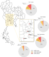
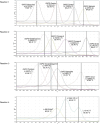
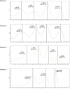
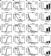
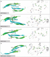
Accurate diagnosis of glucose-6-phosphate dehydrogenase (G6PD) deficiency is crucial for relapse malaria treatment using 8-aminoquinolines (primaquine and tafenoquine), which can trigger haemolytic anaemia in G6PD-deficient individuals. This is particularly important in regions where the prevalence of G6PD deficiency exceeds 3%-5%, including Southeast Asia and Thailand. While quantitative phenotypic tests can identify women with intermediate activity who may be at risk, they cannot unambiguously identify heterozygous females who require appropriate counselling. This study aimed to develop a genetic test for G6PD deficiency using high-resolution melting curve analysis, which enables zygosity identification of 15 G6PD alleles. In 557 samples collected from four locations in Thailand, the prevalence of G6PD deficiency based on indirect enzyme assay was 6.10%, with 8.08% exhibiting intermediate deficiency. The developed high-resolution melting assays demonstrated excellent performance, achieving 100% sensitivity and specificity in detecting G6PD alleles compared with Sanger sequencing. Genotypic variations were observed across four geographic locations, with the combination of c.1311C>T and c.1365-13T>C being the most common genotype. Compound mutations, notably G6PD Viangchan (c.871G>A, c.1311C>T and c.1365-13T>C), accounted for 15.26% of detected mutations. The high-resolution melting assays also identified the double mutation G6PD Chinese-4 + Canton and G6PD Radlowo, a variant found for the first time in Thailand. Biochemical and structural characterisation revealed that these variants significantly reduced catalytic activity by destabilising protein structure, particularly in the case of the Radlowo mutation. The refinement of these high-resolution melting assays presents a highly accurate and high-throughput platform that can improve patient care by enabling precise diagnosis, supporting genetic counselling and guiding public health efforts to manage G6PD deficiency-especially crucial in malaria-endemic regions where 8-aminoquinoline therapies pose a risk to deficient individuals.
Keywords: G6PD deficiency; genotype; high‐resolution melting; mutations; structural stability.
© 2025 The Authors Tropical Medicine & International Health published by John Wiley & Sons Ltd.
Conflict of interest statement
The authors declare no competing interests.
Figures







References
-
- Beutler E. Glucose‐6‐phosphate dehydrogenase: new perspectives. Blood. 1989;73(6):1397–1401. - PubMed
-
- Motulsky AG. Metabolic polymorphisms and the role of infectious diseases in human evolution. 1960. Hum Biol. 1989;61(5–6):835–869; discussion 70–7. - PubMed
-
- Luzzatto L, Ally M, Notaro R. Glucose‐6‐phosphate dehydrogenase deficiency. Blood. 2020;136:1225–1240. - PubMed
-
- Gaetani GF, Rolfo M, Arena S, Mangerini R, Meloni GF, Ferraris AM. Active involvement of catalase during hemolytic crises of favism. Blood. 1996;88(3):1084–1088. - PubMed
MeSH terms
Substances
LinkOut - more resources
Full Text Sources
Medical
Miscellaneous

