. 2025 Mar-Apr;30(2):e70019.
doi: 10.1111/hel.70019.

Flagellar Assembly Factor FliW2 De-Represses Helicobacter pylori FlaA-Mediated Motility by Allosteric Obstruction of Global Regulator CsrA

Affiliations

Flagellar Assembly Factor FliW2 De-Represses Helicobacter pylori FlaA-Mediated Motility by Allosteric Obstruction of Global Regulator CsrA

Marcia Shu-Wei Su et al. Helicobacter. 2025 Mar-Apr.

Abstract

Background: Helicobacter pylori colonizes the human stomach as a dominant member of the gastric microbiota and constitutively expresses flagellar motility for survival. Carbon storage regulator A (CsrA) is a posttranscriptional global regulator and a critical determinant of H. pylori's motility and pathogenicity. The regulation of H. pylori CsrA is still uncertain although in other species CsrA is reported to be antagonized by small RNAs and proteins. In this study, we attempted to unveil how CsrA is regulated and hypothesized that H. pylori CsrA activity is antagonized by a flagellar assembly factor, FliW2, via protein allosteric obstruction.

Materials and methods: Multiple sequence comparisons indicated that, along its length and in contrast to fliW1, the fliW2 of H. pylori J99 is conserved. We then generated an isogenic ΔfliW2 strain whose function was characterized using phenotypic and biochemical approaches. We also applied a machine learning approach (AlphaFold2) to predict FliW2-CsrA binding domains and investigated the FliW2-CsrA interaction using pull-down assays and in vivo bacterial two-hybrid systems.

Results: We observed the reduced expression of major flagellin FlaA and impaired flagellar filaments that attenuated the motility of the ΔfliW2 strain. Furthermore, a direct interaction between FliW2 and CsrA was demonstrated, and a novel region of the C-terminal extension of CsrA was suggested to be crucial for CsrA interacting with FliW2. Based on our AlphaFold2 prediction, this C-terminal region of FliW2-CsrA interaction does not overlap with CsrA's N-terminal RNA binding domain, implying that FliW2 allosterically antagonizes CsrA activity and restricts CsrA's binding to flaA mRNAs.

Conclusions: Our data points to novel regulatory roles that the H. pylori flagellar assembly factor FliW2 has in obstructing CsrA activity, and thus FliW2 may indirectly antagonize CsrA's regulation of flaA mRNA processing and translation. Our findings reveal a new regulatory mechanism of flagellar motility in H. pylori.

Keywords: Helicobacter pylori; CsrA; FliW2; flagellar biosynthesis; major flagellin a (FlaA); motility.


Conflict of interest statement

The authors declare no conflicts of interest.

Figures

FIGURE 1
FIGURE 1
Phylogenetic tree analysis and characteristics of the CsrA and FliW homologs in Proteobacteria. The protein sequences of H. pylori J99 CsrA and its homologs (Table S3) were used to perform phylogenetic tree analysis from a range of representative taxa spanning proteobacteria (left panel) using (standalone) MAFFT v7.526, the L‐INS‐i method and default settings. With the alignment as input, a distance matrix was generated using the WAG amino acid model implemented in phangorn. A tree topology was generated via UPGMA (unweighted pair group method with an arithmetic mean) and also implemented in phangorn. Bootstrap analysis was carried out and the results were plotted using phangorn's plotBS() function. In the right panel, further detail regarding the CsrA and FliW homologs is shown. Not all clades of Proteobacteria possess the carboxyl‐terminal extension of CsrA or show the presence of FliW. Protein homolog comparison was performed using the H. pylori J99 proteins CsrA (UniProt ID: Q9ZJH4), FliW1 (UniProt ID: Q9ZK60), and FliW2 (UniProt ID: Q9ZJL5) as queries for protein identity analysis. It is of note that H. pylori J99 (the third species from the top) possesses the CsrA with the C‐terminal extension and two FliW homologs. The identity of the H. pylori FliW1 is 39.5%, compared to the query, H. pylori FliW2 (100%).
FIGURE 2
FIGURE 2
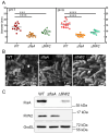
Examination of the motility and flagellation of H. pylori ΔfliW2 strain. (A) Swarming motility examination. H. pylori wild‐type (WT, circle symbol), the non‐motile ΔflaA mutant (square symbol), and the ΔfliW2 mutant (triangle symbol) strains were inoculated on the Brucella soft agar plates at pH 7 (left plot) and pH 6 (right plot). The diameter of bacterial motility was recorded and calculated as the mean ± SD of at least three biological replicates. Error bars are standard deviation. An unpaired t test with Welch's correction was applied to calculate the statistical significance (***p < 0.0005; ****p < 0.0001). (B) Scanning electron microscopic analysis on bacterial morphology and flagellation. Bacterial morphology and flagellar structure were examined in the WT (left plot), ΔflaA (middle plot), and ΔfliW2 (right plot) strains after 55–60 h of inoculation. The formed flagellar filaments are indicated (white arrows). The non‐motile ΔflaA mutant cells were mostly aflagellate, though some possessed short flagella. The micrographs were taken from three fields in two independent experiments. Scale bars represent 1 μm. (C) Expression of major flagellin FlaA by Western blot analysis. Western blot analysis was carried out to examine the expression of FlaA extracted from the whole‐cell proteins of the WT, ΔflaA, and ΔfliW2 strains at the early stationary phase. FlaA and FliW2 were probed using mouse polyclonal antibodies, while a mouse monoclonal antibody was used to detect GroEL. GroEL served as an internal control. The absence of the FlaA and FliW2 signals showed the specificity of anti‐FlaA and anti‐FliW2, respectively.
FIGURE 3
FIGURE 3
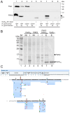
Investigation on CsrA‐FliW2 interaction by pull‐down assays and liquid chromatography with tandem mass spectrometry (LC–MS/MS) analysis. (A) Pull‐down assay using H. pylori lysates. The recombinant polyhistidine‐tagged CsrA protein (CsrAHis) was overexpressed in the E. coli BL21(DE3) host cells, captured, and purified by nickel‐charged magnetic beads under a native condition. Once the CsrAHis‐bound Ni+‐magnetic beads were prepared, we applied the sonication lysate (SL) from the H. pylori wild‐type (WT) cells or the ΔfliW2 cells. After a series of wash steps, the proteins that bound to CsrAHis were co‐eluted and analyzed using Western blotting. The presence of FliW2 signals (~15 kDa, solid triangle) in the eluent of the H. pylori WT lysate and CsrAHis‐bound beads (lane 7) indicated that CsrA interacted with FliW2. The open triangle indicates the non‐specific signal of anti‐FliW2 antibody. In addition to this, in the control group where the H. pylori ΔfliW2 lysate was pooled with CsrAHis, the absence of FliW2 signals (lane 9) confirmed the FliW2‐CsrAHis interaction. Detection of the FlaA protein served as internal control. The data shown was the representative of three independent experiments. TL stands for total lysate, SL for sonication lysate, W for wash fraction, and E for elution fraction. (B) Pull‐down assay using hetero‐expression in the background of E. coli cells. We overexpressed the recombinant CsrAHis (pET22b‐CsrA) (lane 3) and the recombinant tag‐free FliW2 (pET22b‐FliW2‐noHis) (lane 5), respectively, in the E. coli BL21(DE3) host cells. After IPTG induction, the E. coli cell lysates were prepared by sonication and pooled. After pooling, the resulting supernatant was loaded onto a nickel‐NTA affinity column, washed, and the proteins that bound to the nickel column and CsrAHis were eluted. The eluted proteins were examined by SDS‐PAGE and stained. We found the abundant protein bands (15 kDa and 9 kDa) that were presumably tag‐free FliW2 proteins and CsrAHis (lane 7). The data shown was the representative of two biological replicates. M stands for protein marker, NI for non‐induction, I for induction, and E for elution fraction. (C) The validation of CsrA‐bound FliW2 protein by LC–MS/MS analysis. The overexpressed protein band (15 kDa) from Figure 3B (lane 7) was excised and underwent in‐gel trypsin digestion for protein identification using LC–MS/MS analysis. The peptide fragments that matched to those of the FliW2 protein of H. pylori J99 are highlighted in gray in the figure.
FIGURE 4
FIGURE 4
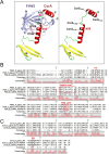
In silico prediction of the FliW2 and CsrA proteins. (A) AlphaFold2 prediction of CsrA‐FliW2 interaction. The three‐dimensional structure of the FliW2‐CsrA complex was predicted using AlphaFold2. The FliW2 protein consists of the β‐sheet regions (left plot, colored lavender), where the previously reported conserved residues for CsrA binding are indicated [38]. CsrA (full length, 1–76 residues) contains the N‐terminal β‐sheet region and a loop–helix–loop–helix region. It should be noted that the C‐terminal extension of CsrA that forms the loop–helix structure might be crucial for FliW2 binding. Therefore, we designed several C‐terminal truncations: CsrA1‐72 (4‐residue deletion), CsrA1‐63 (13‐residue deletion), CsrA1‐58 (18‐residue deletion), and CsrA1‐54 (22‐residue deletion). (B) Protein sequence alignment of the FliW2 homologs. The FliW2 (UniProt ID: Q9ZJL5) of H. pylori strain J99/ATCC 700824, the FliW (UniProt ID: Q0P9H9) of C. jejuni strain ATCC 700819/NCTC 11168, and the FliW (UniProt ID: A0AAE3WUD6) of B. subtilis strain NCIB 3610/ ATCC 6051 were aligned and analyzed. The negative loop and the location of the residues (F27, Q106, V108) that were reported conserved for FliW2 binding to CsrA are labeled. (C) Protein sequence alignment of the CsrA homologs. The CsrA (UniProt ID: Q9ZJH4) of H. pylori strain J99/ATCC 700824, the CsrA (UniProt ID: Q0P9F1) of C. jejuni strain ATCC 700819/NCTC 11168, and the CsrA (UniProt ID: A0AAE3WX71) of B. subtilis strain NCIB 3610/ ATCC 6051 were aligned and analyzed. The location of the conserved active site N55 for CsrA to bind FliW2 is labeled.
FIGURE 5
FIGURE 5
Validation of in vivo CsrA‐FliW2 interaction using a bacterial two‐hybrid system. The Bacterial Adenylate Cyclase‐based Two‐Hybrid (BACTH) system was employed to detect protein interactions by measuring the activity of β‐galactosidase in the E. coli DHM1 strain. In brief, in the first experimental set (upper panel), the fliW2 gene was cloned into pKT25 (N‐terminal T25 fragment fusion; bait vector) to form T25‐FliW2. The full‐length or truncated csrA gene was cloned into pUT18 (C‐terminal T18 fragment fusion; target vector) to form T18‐CsrA. The pKT25‐ and pUT18‐derived plasmids were co‐transformed into the E. coli DHM1 strain. After inoculation with IPTG for 16 h, the cells were extracted for the measurement of their β‐galactosidase activity. In the second experimental set (lower panel), we instead created T25‐CsrA including truncated forms and T18‐FliW2 to examine their protein interactions. An increase of measured β‐galactosidase activity in Miller units is an indication of positive interaction. E. coli DHM1 cells harboring pKT25 and pUT18 served as negative controls and those harboring pKT25‐FeoB and pUT18‐FeoC served as positive controls. The background signal controls (vectors) were included to identify false‐positive interactions. The data shown was the representatives of three biological replicates and calculated as mean ± SD. Error bars are standard deviation (**** p < 0.0001).
FIGURE 6
FIGURE 6
A model of the allosteric obstruction of FliW2 to CsrA that attenuates the effect of CsrA on the motility of H. pylori . In the absence of FliW2 (upper panel), CsrA proteins are free to form dimers. The CsrA dimers bind to either a target transcript itself or its 5′ un‐translational region (UTR), where CsrA motifs (NGGA) are overlapped with ribosomal binding sites (RBSs). Since the RBSs are blocked, ribosomes are unable to initiate the translation of the target transcripts (i.e., major flagellin flaA) and may affect the processing of the target transcripts. This then reduces the expression of FlaA and FlaA‐associated flagellar formation. As a consequence, CsrA modulates the motility of H. pylori through a negative regulatory mechanism at the posttranscriptional level. However, when FliW2 is present (lower panel), FliW2 interacts with CsrA to form heterodimers, thus FliW2 obstructs CsrA by binding to allosteric sites of CsrA at its C‐terminal extension, differing from the RNA binding regions. Therefore, the conformational structures of FliW‐CsrA heterodimers preclude binding to the target transcripts. This allows ribosomes to bind and start the translation of the mRNAs, attenuating the negative regulation of CsrA. Taken together, FliW2 de‐represses the motility of H. pylori by the allosteric obstruction of CsrA.

References

    1. Lei C., Xu Y., Zhang S., Huang C., and Qin J., “The Role of Microbiota in Gastric Cancer: A Comprehensive Review,” Helicobacter 29, no. 2 (2024): e13071. - PubMed
    1. Cheok Y. Y., Lee C. Y. Q., Cheong H. C., et al., “An Overview of Helicobacter pylori Survival Tactics in the Hostile Human Stomach Environment,” Microorganisms 9, no. 12 (2021): 2502. - PMC - PubMed
    1. Ottemann K. M. and Lowenthal A. C., “ Helicobacter pylori Uses Motility for Initial Colonization and to Attain Robust Infection,” Infection and Immunity 70, no. 4 (2002): 1984–1990. - PMC - PubMed
    1. Singh H., Ye A., and Ferrua M. J., “Aspects of Food Structures in the Digestive Tract,” Current Opinion in Food Science 3 (2015): 85–93.
    1. Danielli A., Amore G., and Scarlato V., “Built Shallow to Maintain Homeostasis and Persistent Infection: Insight Into the Transcriptional Regulatory Network of the Gastric Human Pathogen Helicobacter pylori ,” PLoS Pathogens 6, no. 6 (2010): e1000938. - PMC - PubMed

MeSH terms

LinkOut - more resources