Review
. 2025 Jun;85(9):841-849.
doi: 10.1002/pros.24890. Epub 2025 Mar 13.

Incidental Brain Metastases From Prostate Cancer Diagnosed With PSMA PET/CT and MRI: A Case Series and Literature Review

Affiliations
Review

Incidental Brain Metastases From Prostate Cancer Diagnosed With PSMA PET/CT and MRI: A Case Series and Literature Review

Mark Willy L Mondia et al. Prostate. 2025 Jun.

Abstract

Background: Brain metastases (BMETS) from prostate cancer are rare. Hence, brain imaging in neurologically asymptomatic patients with advanced prostate cancer (aPC) is not routinely performed. Prostate-specific membrane antigen (PSMA) PET/CT uses a radiotracer that binds to prostate cancer epithelial cells and is FDA-approved for initial staging for high-risk prostate cancer, detecting prostate cancer recurrence, and determining eligibility for radionuclide therapy.

Methods: We report six patients with asymptomatic BMETS from aPC found on staging PSMA PET/CT or MRI. Along with cranial MRI, PSMA PET/CT may be useful for detecting asymptomatic intracranial metastasis in select patients with prostate cancer.

Results: Brain metastases were diagnosed in four patients by staging PSMA PET/CT scan-three after systemic disease progression and one during routine surveillance. In two other patients, BMETS were detected using MRI despite negative PSMA PET/CT for brain lesions. All were neurologically asymptomatic. Three patients had undetectable serum prostate-specific antigen (PSA) concentrations; one had neuroendocrine differentiation on histology.

Conclusion: In patients with poorly differentiated or neuroendocrine aPC, BMETS may occur without neurologic symptoms and stable PSA. PSMA PET/CT may complement brain MRI for identifying BMETS in these patients.

Keywords: MRI; PET/CT; PSMA; brain metastasis; prostate cancer.


Conflict of interest statement

The authors declare no conflicts of interest.

Figures
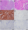
Figure 1
Figure 1
Histopathologic findings for Case 6. Low power view (100× objective) of hematoxylin and eosin (H&E) stained section (A), illustrating compact, solid growth pattern. High power view (400×) of H&E section (B), illustrating pleomorphic epithelioid cells with vacuolated cytoplasm and prominent nucleoli. High power view (400×) of positive keratin cocktail stain (C). High powered view (400×) of positive staining for NKX3.1 (D) in a subset of tumor cell nuclei. No staining for synaptophysin (E) or chromogranin (F). Scale bar: 200 µM in (A), 50 µM in (B–F). [Color figure can be viewed at wileyonlinelibrary.com]
Figure 2
Figure 2
Radiologic findings for Case 2. A 68‐year‐old man with metastatic prostate cancer. (A) Axial PSMA PET, and (B) Axial fused PET/CT revealed a 1.5 cm focus of mild uptake (SUV max 6.1) in the left parietal lobe (arrows). (C and D) Axial post‐contrast T1WI from a follow‐up MRI shows a heterogeneous enhancing lesion in the left parietal lobe (two arrows). An additional punctate enhancing lesion is seen in the left centrum semiovale (single arrow) without a corresponding radiotracer avid focus on the previous PSMA PET. This is likely due to background noise. However, this focus was identified in subsequent PSMA PET (not shown). [Color figure can be viewed at wileyonlinelibrary.com]
Figure 3
Figure 3
Radiologic findings for Case 5. A 67‐year‐old man with castration‐resistant metastatic prostatic carcinoma. (A and B) Axial PSMA PET, and (C and D) Corresponding axial fused PET/CT (coverage was from skull base to upper thighs) show radiotracer avid lesions (red arrows) in the left posterior temporal lobe (1 cm size, SUV max 5.4, intermediate PSMA expression) and right cerebellum (5 mm size, SUV max 2.5, low PSMA expression) consistent with prostate cancer metastasis. (E–H) Axial post‐contrast T1WI done 2 weeks after the PSMA PET/CT shows corresponding enhancing lesions (red arrows). However, there are additional enhancing < 5 mm size metastatic lesions in the left cerebellum and left parietal parasagittal cortex. This discrepancy is likely due to the limited resolution of these small lesions on PSMA PET/CT and the lack of coverage of the left parietal lesion. [Color figure can be viewed at wileyonlinelibrary.com]
Figure 4
Figure 4
Radiologic findings for Case 6. A 62‐year‐old man with metastatic prostatic carcinoma screened for brain metastasis as part of surveillance. (A and B) Axial PET, and fused PET/CT showing an incidental 4 cm left parietal lobe mass (arrows) with significant PSMA expression (SUV max 7.8). (C) Axial post‐contrast T1WI showing a corresponding enhancing mass (arrow). [Color figure can be viewed at wileyonlinelibrary.com]

References

    1. Siegel R. L., Miller K. D., Wagle N. S., and Jemal A., “Cancer Statistics, 2023,” CA: A Cancer Journal for Clinicians 73, no. 1 (2023): 17–48, 10.3322/caac.21763. - DOI - PubMed
    1. Gandaglia G., Abdollah F., Schiffmann J., et al., “Distribution of Metastatic Sites in Patients With Prostate Cancer: A Population‐Based Analysis,” Prostate 74, no. 2 (2014): 210–216, 10.1002/pros.22742. - DOI - PubMed
    1. Sutton M. A., Watkins H. L., Green L. K., and Kadmon D., “Intracranial Metastases as the First Manifestation of Prostate Cancer,” Urology 48, no. 5 (1996): 789–793, 10.1016/S0090-4295(96)00238-5. - DOI - PubMed
    1. Bubendorf L., Schöpfer A., Wagner U., et al., “Metastatic Patterns of Prostate Cancer: An Autopsy Study of 1,589 Patients,” Human Pathology 31, no. 5 (2000): 578–583, 10.1053/hp.2000.6698. - DOI - PubMed
    1. McLaughlin L. A., Yildirim O., Rosenblum M. K., et al., “Identification of Incidental Brain Tumors in Prostate Cancer Patients via PSMA PET/CT,” Journal of Neuro‐Oncology 163, no. 2 (2023): 455–462, 10.1007/s11060-023-04355-x. - DOI - PMC - PubMed

MeSH terms