. 2025 Jun;116(6):1773-1784.
doi: 10.1111/cas.70038. Epub 2025 Mar 13.

Claudin-11 Enhances Invasive and Metastatic Abilities of Small-Cell Lung Cancer Through MT1-MMP Activation

Affiliations

Claudin-11 Enhances Invasive and Metastatic Abilities of Small-Cell Lung Cancer Through MT1-MMP Activation

Shuichi Sakamoto et al. Cancer Sci. 2025 Jun.

Abstract

Small-cell lung cancer (SCLC) is an aggressive tumor characterized by the frequent development of distant metastases. This study aimed to explore the mechanism of SCLC metastasis using an originally developed orthotopic transplantation model with DMS273 cells. An analysis of G3H cells, a highly metastatic subline of DMS273 cells, revealed that claudin-11 promotes the invasive and metastatic ability of the cells. Further analysis revealed that membrane type 1-matrix metalloproteinase (MT1-MMP), which degrades a wide range of extracellular matrix components, was coprecipitated with claudin-11. Gelatin zymography revealed that claudin-11 enhanced MT1-MMP activity, and MT1-MMP silencing suppressed the invasive and metastatic ability of G3H cells. Moreover, in MT1-MMP silencing DMS273 cells, the enhancement of invasion and metastatic potential induced by CLDN11 overexpression was abolished. These results demonstrate that claudin-11 enhances the invasive capacity of the cells by activating MT1-MMP, which promotes metastatic formation in the orthotopic transplantation model. Additionally, claudin-11 expression was detected in SCLC tumor samples, and higher expression of CLDN11 correlated with poor prognosis in patients with SCLC. These findings suggest that the claudin-11/MT1-MMP axis plays an important role in SCLC pathogenesis.

Keywords: MT1‐MMP; SCLC; claudin‐11; invasion; metastasis.


Conflict of interest statement

Masanori Hatakeyama is the Editor‐in‐Chief of Cancer Science. The others have no conflicts of interest.

Figures

FIGURE 1
FIGURE 1
Gene expression profile of a highly metastatic subline G3H. (a) Formation of metastatic tumors from the highly metastatic subline G3H and parental DMS273‐GFP in the orthotopic model. (b) Matrigel invasive ability of G3H and DMS273‐GFP assessed using the transwell chamber assay. (c) Volcano plot of the G3H and DMS273‐GFP gene expression profiles determined using mRNA microarray analysis. (d) Scatter plot showing the significantly expressed genes between G3H and DMS273‐GFP cells (Fold change > 3; p < 0.01) determined using mRNA microarray analysis.
FIGURE 2
FIGURE 2
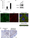
Claudin‐11 expression in the highly metastatic subline G3H and parental DMS273‐GFP. (a) RT‐qPCR analysis of CLDN11 mRNA in DMS273‐GFP and G3H cells. Total RNA (0.5 μg) was analyzed. Data are presented as mean + standard deviation (SD) of three independent experiments performed in duplicate. (b) Western blot analysis of claudin‐11 in DMS273‐GFP and G3H cells. (c) Representative immunocytochemical images with claudin‐11 antibodies in DMS273‐GFP and G3H. Scale bar: 20 μm. (d) Representative immunohistochemistry images of claudin‐11 antibodies in the lung and metastatic tumors of the orthotopic metastatic model using G3H. Scale bars: 50 μm.
FIGURE 3
FIGURE 3
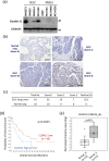
Expression of claudin‐11 in human SCLC cell lines and lung tumors of SCLC patients. (a) Western blot analysis of claudin‐11 in human lung cancer cell lines. (b) Representative images of the tissue microarray analysis of lung tumors of patients with SCLC with claudin‐11 antibodies. A human SCLC tissue microarray (BS04116, US Biomax) was used. Scale bars: 100 μm. (c) Summary of the tissue microarray analysis with claudin‐11 antibodies. (d) Kaplan–Meier plot of overall survival in patients with SCLC with high (n = 41) versus low (n = 36) expression of CLDN11 mRNA expression (Log‐rank test p = 0.0425). (e) Box plot for CLDN11 mRNA expression levels based on expression array data of 23 patients with SCLC from GSE43346.
FIGURE 4
FIGURE 4
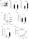
CLDN11 overexpression enhances the invasive ability and metastatic tumor formation of DMS273‐GFP cells. (a) Western blot analysis of claudin‐11 in DMS273‐GFP cells transfected stably with a CLDN11 expression lentivirus vector (CLDN11) or a control vector (Vec). (b) In vitro growth rate for 72 h of CLDN11‐transfected (CLDN11) and control cells (Vec) determined using the MTT assay. Data are presented as mean ± SD. (c) Invasive abilities of CLDN11‐transfected (CLDN11) and control cells (Vec) determined using the Matrigel Transwell chamber assay. Data are presented as mean ± SD. (d, e) Orthotopic and metastatic tumor formations in the orthotopic transplantation model with CLDN11‐transfected cells (CLDN11) and control cells (Vec). 2.5 × 105 cells were transplanted into the left lung of each nude mouse. Mice were sacrificed when they became moribund, and orthotopic and metastatic tumors were evaluated. (d) Distant metastatic tumor formation in the model. Dotted lines indicate the mean number of organs positive for metastasis in each mouse. Percentages indicate the incidence of distant metastases. (e) Orthotopic tumor formation. Results are expressed as mean ± SD. (f) Formation of metastatic colonies in lungs in experimental metastasis models generated using CLDN11‐transfected (CLDN11) and control cells (Vec). 1 × 106 cells were injected into the tail vein of each nude mouse (n = 10). Mice were sacrificed 7 weeks after inoculation, and metastatic tumor foci in the lungs were assessed. Results are expressed as mean ± SD. (g) Subcutaneous tumor growth of CLDN11‐transfected (CLDN11) and control cells (Vec). 1 × 106 cells were subcutaneously inoculated into nude mice (n = 6). Tumor volumes are presented as mean ± SD.
FIGURE 5
FIGURE 5
CLDN11 suppression decreases the invasive ability and metastatic tumor formation of G3H cells. (a) RT‐qPCR analysis of CLDN11 mRNA in CLDN11‐silenced G3H (shCLDN11#1, shCLDN11#2), control shRNA‐transduced G3H (shNC) and parental cells (G3H). Total RNA (0.5 μg) was subjected to analysis. Data are presented as mean + SD. (b) Western blot analysis of claudin‐11 in CLDN11‐silenced G3H (shCLDN11#1, shCLDN11#2), control shRNA transduced G3H (shNC) and parental cells (G3H). (c) In vitro growth rate for 72 h of CLDN11‐silenced G3H (shCLDN11#1, shCLDN11#2), control shRNA transduced G3H (shNC) and parental cells (G3H) determined by MTT assay. Data are presented as mean + SD. (d) Invasive abilities of CLDN11‐silenced G3H (shCLDN11#1, shCLDN11#2) and control shRNA‐transduced G3H cells (shNC) determined using the Transwell chamber assay. Data are expressed as mean + SD. (e, f) Orthotopic and metastatic tumor formation in the orthotopic transplantation model with CLDN11‐silenced G3H (shCLDN11#1) and G3H control cells transduced with shRNA (shNC). 2.5 × 105 cells were transplanted into the left lung of each nude mouse. (e) Distant metastatic tumor formation in the model. Dotted lines indicate the mean number of organs positive for metastasis in each mouse. Percentages show the incidence of distant metastases. (f) Orthotopic tumor formation. Results are expressed as mean + SD.
FIGURE 6
FIGURE 6
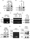
CLDN11 stimulates proMMP2 processing in DMS273 cells and HEK293T cells. (a) Immunoprecipitation (IP) with a FLAG antibody in DMS273‐GFP cells stably transfected with a 3 × FLAG‐tagged CLDN11 expression lentivirus vector (CLDN11) or a control vector (Vec). (b) IP with a MT1‐MMP antibody or a control Rabbit IgG in G3H cells. (c) Gelatin zymography analysis of DMS273‐GFP cells stably transfected with a 3 × FLAG‐tagged CLDN11 expression lentivirus vector (CLDN11) or a control vector (Vec). (d) Gelatin zymography analysis of CLDN11‐silenced G3H cells (shCLDN11#1, shCLDN11#2) and control shRNA‐transduced G3H cells (shNC). (e) Western blot analysis of MT1‐MMP in CLDN11‐silenced G3H (shCLDN11#1, shCLDN11#2) and control shRNA‐transduced G3H (shNC). (f) IP with a FLAG antibody of HEK293T cells cotransfected with HA‐CLDN11 and FLAG‐tagged wild‐type MT1‐MMP plasmids. (g) Gelatin zymography analysis of HEK293T cells transiently transfected with CLDN11 and MT1‐MMP. An HA‐CLDN11 plasmid was cotransfected with an MT1‐MMP‐FLAG plasmid into HEK293T cells. The cells were lysed 48 h after transfection, and the lysates were analyzed. (h) Cell surface biotinylation assay. HEK293T cells were transiently transfected with CLDN11 and MT1‐MMP and incubated with EZ‐Link Sulfo‐NHS‐Biotin 24 h after transfection. The cells were lysed and immunoprecipitated with anti‐FLAG antibody. The IP products were separated by SDS‐PAGE. The biotinylated proteins were detected using streptavidin‐HRP.
FIGURE 7
FIGURE 7
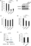
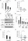
Role of the C‐terminus domain of claudin‐11 in the interaction with MT1‐MMP. (a) Invasive abilities of MT1‐MMP‐silenced G3H cells (shMT1‐MMP#1, shMT1‐MMP#2) and control shRNA‐transduced G3H cells (shNC) determined using the Transwell chamber assay. Data are presented as mean + SD. (b) Distant metastatic tumor formation in the orthotopic transplantation model with MT1‐MMP‐silent G3H cells (shMT1‐MMP#2) and G3H control cells transduced with shRNA (shNC). 2.5 × 105 cells were transplanted into the left lung of each nude mouse. Dotted lines indicate the mean number of organs positive for metastasis in each mouse. Percentages show the incidence of distant metastases. (c) Western blot analysis of MT1‐MMP and claudin‐11 in DMS273‐GFP cells overexpressing 3 × FLAG‐tagged wild‐type CLDN11 with MT1‐MMP silencing (shMT1‐MMP#1, shMT1‐MMP#2) and control shRNA (shNC). (d) Invasive abilities of DMS273‐GFP cells overexpressing 3 × FLAG‐tagged wild‐type CLDN11 with MT1‐MMP silencing (shMT1‐MMP#1, shMT1‐MMP#2) and control shRNA (shNC). Data are expressed as mean + SD. (e, f) Orthotopic and metastatic tumor formation in the orthotopic transplantation model with DMS273‐GFP cells overexpressing CLDN11 with MT1‐MMP silencing (shMT1‐MMP#2). 2.5 × 105 cells were transplanted into the left lung of each nude mouse. Dotted lines indicate the mean number of organs positive for metastasis in each mouse. Percentages show the incidence of distant metastases.
FIGURE 8
FIGURE 8
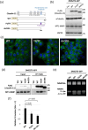
Role of the C‐terminus domain of claudin‐11 in the stimulation of MT1‐MMP. (a) Schematic of the C‐terminus cytoplasmic domain mutants of human claudin‐11. (b) Western blot analysis of 3 × FLAG‐tagged wild‐type CLDN11 (WT), Y191F/Y192F‐CLDN11 mutant (YY/FF), del191‐CLDN11 mutant (del191‐) and MT1‐MMP in DMS273‐GFP cells stably transfected with expression lentivirus vectors of the claudin‐11 mutants. (c) Representative immunocytochemical images with a FLAG antibody of the claudin‐11 mutants in DMS273‐GFP cells. FLAG in green, nuclei in blue. Scale bars: 25 μm. (d) IP with a FLAG antibody from DMS273‐GFP cells stably transfected with the CLDN11 mutants. (e) Gelatin zymography analysis of DMS273‐GFP cells stably transfected with the CLDN11 mutants. (f) Invasive abilities of DMS273‐GFP cells stably transfected with the CLDN11 mutants determined using the Transwell chamber assay. Data are presented as mean + SD.

References

    1. Gazdar A. F., Bunn P. A., and Minna J. D., “Small‐Cell Lung Cancer: What We Know, What We Need to Know and the Path Forward,” Nature Reviews Cancer 17, no. 12 (2017): 725–737, 10.1038/nrc.2017.87. - DOI - PubMed
    1. Sabari J. K., Lok B. H., Laird J. H., Poirier J. T., and Rubin C. M., “Unravelling the Biology of SCLC: Implications for Therapy,” Nature Reviews Clinical Oncology 14, no. 9 (2017): 549–561, 10.1038/nrclinonc.2017.71. - DOI - PMC - PubMed
    1. Ko J., Winslow M. M., and Sage J., “Mechanisms of Small Cell Lung Cancer Metastasis,” EMBO Molecular Medicine 13, no. 1 (2021): e13122, 10.15252/emmm.202013122. - DOI - PMC - PubMed
    1. Sakamoto S., Inoue H., Ohba S., et al., “New Metastatic Model of Human Small‐Cell Lung Cancer by Orthotopic Transplantation in Mice,” Cancer Science 106, no. 4 (2015): 367–374, 10.1111/cas.12624. - DOI - PMC - PubMed
    1. Sakamoto S., Inoue H., Kohda Y., Ohba S. I., Mizutani T., and Kawada M., “Interferon‐Induced Transmembrane Protein 1 (IFITM1) Promotes Distant Metastasis of Small Cell Lung Cancer,” International Journal of Molecular Sciences 21, no. 14 (2020): 4934, 10.3390/ijms21144934. - DOI - PMC - PubMed

MeSH terms

LinkOut - more resources