. 2025 Mar 13;10(8):e179875.
doi: 10.1172/jci.insight.179875. eCollection 2025 Apr 22.

Tape strip expression profiling of juvenile dermatomyositis skin reveals mitochondrial dysfunction contributing to disease endotype

Affiliations

Tape strip expression profiling of juvenile dermatomyositis skin reveals mitochondrial dysfunction contributing to disease endotype

Jessica L Turnier et al. JCI Insight. .

Abstract

Skin inflammation in juvenile dermatomyositis (JDM) can signal disease onset or flare, and the persistence of cutaneous disease can prevent complete disease remission. The noninvasive study of cutaneous expression signatures through tape stripping (TS) holds the potential to reveal mechanisms underlying disease heterogeneity and organ-specific inflammation. The objectives of this study were to (a) define TS expression signatures in lesional and nonlesional JDM skin, (b) analyze TS signatures to identify JDM disease endotypes, and (c) compare TS and blood signatures. Although JDM lesional skin demonstrated interferon signaling as the top upregulated pathway, nonlesional skin uniquely highlighted pathways involved in metabolism, angiogenesis, and calcium signaling. Both lesional and nonlesional skin shared inflammasome pathway dysregulation. Using unsupervised clustering of skin expression data, we identified a treatment-refractory JDM subgroup distinguished by upregulation of genes associated with mitochondrial dysfunction. The treatment-refractory JDM subgroup also demonstrated higher interferon, angiogenesis, and innate immune expression scores in skin and blood, though scores were more pronounced in skin as compared with blood. TS expression signatures in JDM provided insight into disease mechanisms and molecular subgroups. Skin, as compared with blood, transcriptional profiles served as more sensitive markers to classify disease subgroups and identify candidate treatment targets.

Keywords: Autoimmune diseases; Autoimmunity; Clinical practice; Dermatology; Expression profiling; Inflammation.


Figures
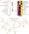
Figure 1
Figure 1. Biological signatures identified in JDM L and NL skin compared with CTL.
(A) The left panel displays the top 10 biological pathways (P value < 0.05) regulated in JDM L (n = 17) and NL skin (n = 28) compared with CTL skin (n = 20); the right panel displays selected genes from relevant pathways. Canonical pathway P values were computed by the Ingenuity Pathway Analysis (IPA; QIAGEN) software. nNOS, neuronal nitric oxide synthase. (B) Literature-based network analysis of shared and unique genes in JDM L and NL compared with control skin.
Figure 2
Figure 2. Comparison of tape stripping with full-thickness skin biopsy expression signatures.
(A) JDM lesional skin. FDR was extracted from the Limma analysis (B) A 23-gene signature from overlap genes in L expression datasets. Data are presented as mean ± SD. Unpaired 2-tailed parametric Student’s t test with Benjamini-Hochberg correction was used for the comparison between 2 groups. (C) JDM NL skin. FDR was extracted from the Limma analysis. (D) A 100-gene signature from overlap genes in NL expression datasets. Data are presented as mean ± SD. Unpaired 2-tailed parametric Student’s t test with Benjamini-Hochberg correction was used for the comparison between 2 groups. RNA-Seq: n = 20 CTL, n = 17 JDM_L, n = 28 JDM_NL; microarrays: n = 8 CTL, n = 9 JDM_L, n = 6 JDM_NL.
Figure 3
Figure 3. Unsupervised hierarchical clustering of skin expression data identifies subgroups of patients with JDM.
(A) Heatmap of selected DEGs representing pathways reflecting 2 JDM skin subgroups. (B) JDM subgroup 2 demonstrated higher skin-directed interferon, mitochondrial dysfunction, angiogenesis, and innate immune expression scores in skin compared with subgroup 1 and healthy controls. (C) The NFE2L2 signature score was higher in subgroup 2 compared with subgroup 1 and positively associated with the skin-directed interferon score. Unpaired 2-tailed parametric Student’s t test with Benjamini-Hochberg correction was used for the comparison between 2 groups. Cluster 1: n = 65 total samples (n = 45 JDM_NL and 20 JDM_L) corresponding to n = 28 patients; cluster 2: n = 12 total samples (n = 10 JDM_NL and 2 JDM_L) corresponding to n = 8 patients.
Figure 4
Figure 4. Validation of a JDM subgroup expression signature using a JDM skin biopsy microarray dataset.
(A) Heatmap of the DEGs representing selected pathways commonly activated between the 2 identified JDM subgroups in both skin tape stripping and biopsy. (B) Subgroup 2 demonstrated higher mitochondrial/oxidative phosphorylation dysfunction expression scores in skin compared with subgroup 1 and healthy controls. (C) Comparison of JDM skin expression signatures from tape stripping and full-thickness skin biopsies between the 2 identified subgroups. Unpaired 2-tailed parametric Student’s t test with Benjamini-Hochberg correction was used for the comparison between 2 groups. Cluster 1 RNA-Seq: n = 45 JDM_NL and 20 JDM_L samples corresponding to n = 28 patients; cluster 2 RNA-Seq: n = 10 JDM_NL and 2 JDM_L samples corresponding to n = 8 patients; cluster 1 microarrays: n = 6 CTL, 6 JDM_NL and 3 JDM_L samples/patients; cluster 2 microarrays: n = 0 JDM_NL and 6 JDM_L samples/patients.
Figure 5
Figure 5. Whole blood as compared with skin transcriptional profile to define subgroups.
(A) Blood expression scores for pathways of interest. Patients with both NL and L skin samples that were split between the 2 defined molecular subgroups are represented by unfilled circles. The blood samples were assigned to subgroup 2 for analysis. (B) Top 10 canonical pathways from the DEGs in L skin from JDM patients with lower Manual Muscle Testing scores (MMT-8 ≤ 146) compared with controls (FDR < 0.10). Unpaired 2-tailed parametric Student’s t test with Benjamini-Hochberg correction was used for the comparison between 2 groups. CTL: n = 6; cluster 1: n = 34 JDM samples; cluster 2: n = 9 JDM samples. Canonical pathway P values were computed by the IPA software.
Figure 6
Figure 6. Immune cell enrichment analysis in JDM and control skin using CIBERSORTx.
(A) Heatmap from each relevant immune cell type relative fraction in CTL (n = 21), NL (n = 55) JDM, and L (n = 22) JDM skin. (B) Graphs illustrating the relative fraction of B cells, plasma cells, macrophages, dendritic cells, and neutrophils as computed by CIBERSORTx in CTL and JDM samples (each dot represents 1 sample). (C) Heatmap from each relevant immune cell type relative fraction in each sample from control skin and samples in each identified skin subgroup. (D) Graphs illustrating the relative fraction of B cells and macrophages as computed by CIBERSORTx in each CTL and JDM subgroup sample (each dot represents 1 sample). CTL: n = 21, JDM_NL: n = 55, JDM_L: n = 22, JDM subgroup 1: n = 65, JDM subgroup 2: n = 12. Unpaired 2-tailed parametric Student’s t test with Benjamini-Hochberg correction was used for the comparison between 2 groups.

References

    1. Lundberg IE, et al. 2017 European League Against Rheumatism/American College of Rheumatology classification criteria for adult and juvenile idiopathic inflammatory myopathies and their major subgroups. Arthritis Rheumatol. 2017;69(12):2271–2282. doi: 10.1002/art.40320. - DOI - PMC - PubMed
    1. Almeida B, et al. Analysis of published criteria for clinically inactive disease in a large juvenile dermatomyositis cohort shows that skin disease is underestimated. Arthritis Rheumatol. 2015;67(9):2495–2502. doi: 10.1002/art.39200. - DOI - PMC - PubMed
    1. Wang A, et al. Skin disease is more recalcitrant than muscle disease: A long-term prospective study of 184 children with juvenile dermatomyositis. J Am Acad Dermatol. 2021;84(6):1610–1618. doi: 10.1016/j.jaad.2020.12.032. - DOI - PubMed
    1. Mathiesen P, et al. Long-term outcome in patients with juvenile dermatomyositis: a cross-sectional follow-up study. Scand J Rheumatol. 2012;41(1):50–58. - PubMed
    1. Stringer E, et al. Predicting the course of juvenile dermatomyositis: significance of early clinical and laboratory features. Arthritis Rheum. 2008;58(11):3585–3592. doi: 10.1002/art.23960. - DOI - PubMed

LinkOut - more resources