. 2025 Apr 3;112(4):829-845.
doi: 10.1016/j.ajhg.2025.02.016. Epub 2025 Mar 12.

Bi-allelic MED16 variants cause a MEDopathy with intellectual disability, motor delay, and craniofacial, cardiac, and limb malformations

Affiliations

Bi-allelic MED16 variants cause a MEDopathy with intellectual disability, motor delay, and craniofacial, cardiac, and limb malformations

Charlotte Guillouet et al. Am J Hum Genet. .

Abstract

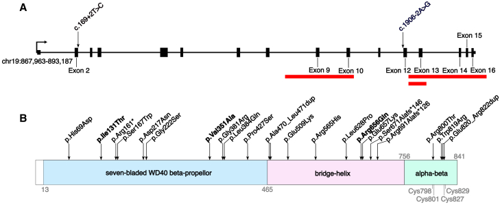
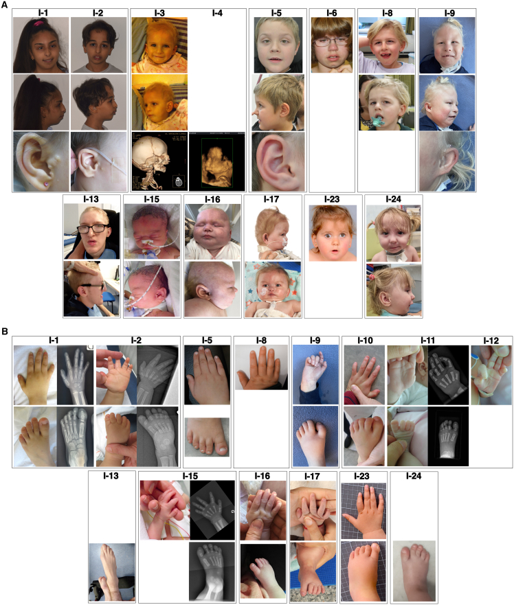
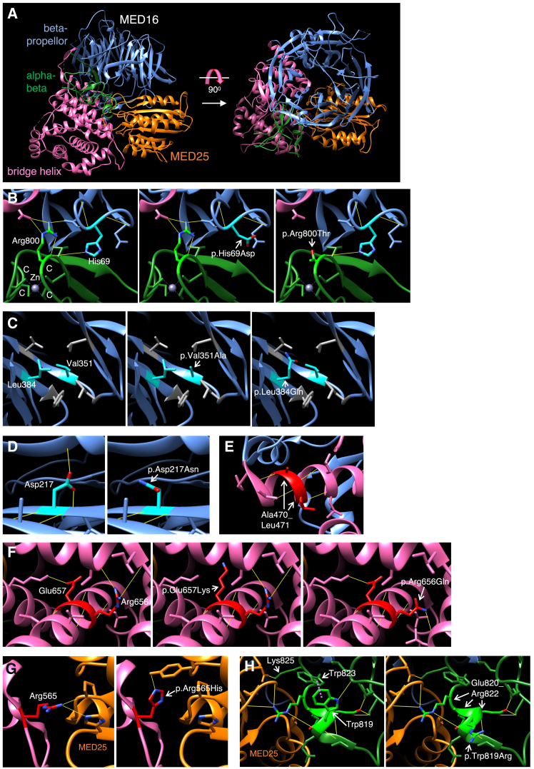
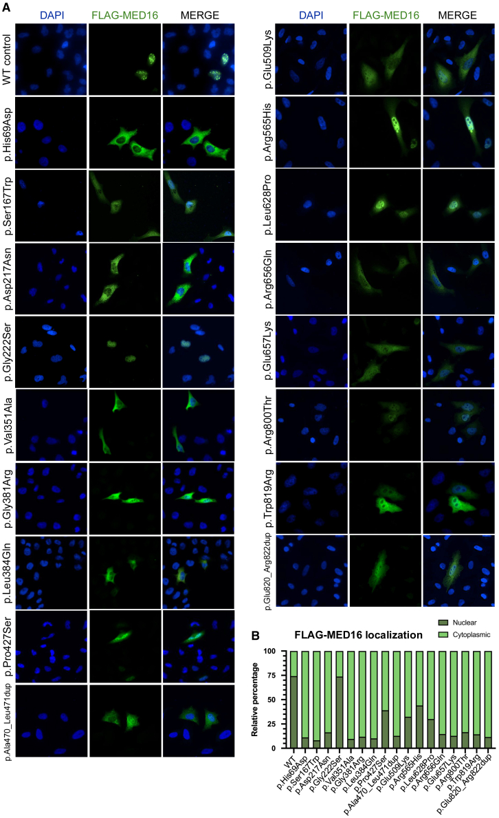
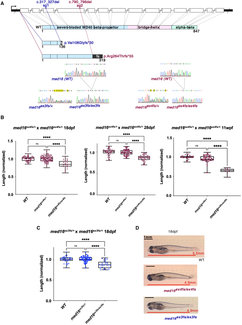
The Mediator complex regulates protein-coding gene transcription by coordinating the interaction of upstream enhancers with the basal transcription machinery at the promoter. Pathogenic variants in Mediator subunits typically lead to neurodevelopmental or neurodegenerative disorders with variable clinical presentations, designated as MEDopathies. Here, we report the identification of 25 individuals from 18 families with bi-allelic MED16 variants who have a multiple congenital anomalies (MCAs)-intellectual disability syndrome. Intellectual disability, speech delay, and/or motor delay of variable severity were constant and associated with variable combinations of craniofacial defects (micro/retrognathia, cleft palate, and preauricular tags), anomalies of the extremities, and heart defects (predominantly tetralogy of Fallot). Visual impairment, deafness, and magnetic resonance imaging (MRI) abnormalities were also frequent. The 26 variants identified were comprised of eight predicted protein-truncating (three intragenic deletions, two frameshifts, and one nonsense and two essential splice site variants) and 18 missense or in-frame duplication variants affecting conserved residues, without clear correlation between phenotypic severity and variant type combination. Three-dimensional modeling indicated that the missense and duplication variants likely have a destabilizing effect on the structural elements of the protein. Immunofluorescence assays demonstrated protein mislocalization from the nucleus to the cytoplasm for 16 of the 17 variants studied. Homozygous mutant med16 zebrafish presented growth delay and increased mortality compared with wild-type fish, and Med16 knockout mice are preweaning lethal, highlighting the conserved requirement of MED16 for development. Overall, we describe an autosomal recessive MCAs-intellectual disability MEDopathy, emphasizing the importance of Mediator during neurodevelopment and suggesting that some tissues are particularly sensitive to the loss of certain subunits.

Keywords: MED16; MEDopathies; Mediator complex; multiple congenital anomalies-intellectual disability syndrome.


Conflict of interest statement

Declaration of interests The authors declare no competing interests.

Figures

Figure 1
Figure 1
Distribution of pathogenic MED16 variants (A) Deletions and variants affecting essential splice sites. Red bars indicate the deletions. Exons and introns are represented according to the MANE transcript GenBank: NM_005481.3, and genomic coordinates are for Hg38. (B) Variants affecting the coding sequence. Bold, the recurrent variants; blue, seven-bladed WD40 beta-propellor; pink, bridge helix; green, alpha-beta domain. The cysteines of the C2C2 zinc finger are indicated in gray beneath the protein. Domain boundaries are based on the crystal structure in Chen et al. (PDB: 7EMF).
Figure 2
Figure 2
Clinical features of the face and extremities in the MED16-associated syndrome Individual (I) numbers are shown at the top of each image. (A) Recurrent facial features shown include micro- and/or retrognathia (I-1, I-2, I-3, I-4, I-6, I-9, I-15, I-17, and I-24), malar hypoplasia (I-1, I-2, and I-3), preauricular tags (I-1, I-2, I-3, and I-15), and puffy eyelids (I-8, I-9, I-15, and I-17). (B) Recurrent hand and feet abnormalities shown include thumb hypoplasia (I-1, I-2, I-9, I-15, and I-17) or aplasia (I-11), 5th finger hypoplasia (I-1, I-2, I-11, and I-15), and hypoplastic fingernails (I-1, I-9, I-10, I-15, and I-17) or toenails (I-1, I-2, I-9, I-10, I-11, and I-17).
Figure 3
Figure 3
Three-dimensional structure of MED16 and modeling of missense variants (A) Structure of MED16 (PDB: 7EMF) with subdomains as defined in Chen et al.: beta-propellor (blue), bridge helix (pink), and alpha-beta domain (green). MED25 (in orange) is the only other Mediator subunit depicted, given its relevance to MED16 variants studied here. Right: view of the upper surface of the MED16 beta-propellor. (B–H) Wild-type and variant amino acids falling in the above domains are colored cyan, red, and bright green, respectively, with hydrogen bonds represented by yellow lines. (B) Left: interdomain interactions mediated by His69 and Arg800. The four cysteines of the C2C2 zinc finger, coordinating a zinc ion (Zn), are each indicated by a “C.” Middle and right: loss of hydrogen bonding for the mutations p.His69Asp and p.Arg800Thr, respectively. (C) Left: Val351 and Leu384 in close proximity within the beta-propellor. Other hydrophobic residues near Val351 and Leu384 are shown in gray. Middle and right, the mutations p.Val351Ala and p.Leu384Gln, respectively. (D) Loss of hydrogen bonding upon mutation of Asp217 (left) to Asn (right). (E) Ala470-Leu471, duplicated in individuals 19 and 20, fall within a helix at the beginning of the bridge helix. The duplication itself is not modeled here. (F) Disrupted hydrogen bonding for the adjacent variants p.Arg656Gln (right) and p.Glu657Lys (middle) within the bridge helix. (G) MED16-MED25 interaction mediated by Arg565 hydrogen bonding (left) is disrupted upon mutation to His (right). (H) Left: Trp819 to Arg822 mediate interactions within the alpha-beta domain and with MED25. Right: p.Trp819Arg mutation disrupts hydrogen bonding and a pi-stacking Trp819-Trp823 interaction (manually drawn as a violet dotted line on left, based on the depiction of the structure in the RCSB Protein Data Bank). Duplication of Glu820_Arg822 in individual 16 would disrupt interactions involving Trp823 and Lys825 immediately downstream.
Figure 4
Figure 4
Subcellular localization of MED16 protein harboring variants Immunofluorescence analysis was performed on HeLa cells transiently transfected with wildtype or mutant FLAG-MED16 expression plasmids. (A) Representative cells for each plasmid (20× magnification). Blue, DAPI; green, FLAG-MED16. WT, wild type. (B) Relative percentage of FLAG-positive cells with nuclear FLAG-MED16 localization in dark green and cytoplasmic FLAG-MED16 localization in light green, for each variant, ordered N- to C-terminally. A cell was scored as cytoplasmic if any degree of specific fluorescence was observed in the cytoplasm, even if nuclear fluorescence was also observed in the same cell. Mean number of FLAG-positive cells analyzed per variant: 199 (range: 166–244). Three independent transfections were performed for each plasmid.
Figure 5
Figure 5
Characterization of med16 mutant zebrafish (A) Schematic of zebrafish med16, with exons depicted as white boxes and untranslated regions as black boxes. Med16 WT and truncated proteins are depicted below the gene: blue, seven-bladed WD40 beta-propellor; pink, bridge helix; green, alpha-beta domain. The single guide RNA sg1, targeting exon 3, induced an 11 bp deletion, resulting in a truncated protein (c.317_327del [GenBank: NM_001199872] [p.Val106Glyfs30]; line med16ex3fs; dark blue text). sg2, targeting exon 4, induced a 7 bp deletion and a truncated protein (c.790_796del [GenBank: NM_001199872] [p.Arg264Thrfs55]; line med16ex4fs; dark red text). Chromatograms show the region of the deletions in WT, heterozygous, and homozygous fish from med16ex3fs and med16ex4fs lines. (B) med16ex4fs/ex4fs fish show growth delay at 18 dpf, 28 dpf, and 11 wpf compared to heterozygotes and WTs. Length was normalized to the mean length of the WT individuals in each tank before pooling genotypes. Total numbers of progeny measured at 18 dpf: WT = 47, med16ex4fs/+ = 88, and med16ex4fs/ex4fs = 25; at 28 dpf: WT = 46, med16ex4fs/+ = 82, and med16ex4fs/ex4fs = 48; at 11 wpf: WT = 30, med16ex4fs/+ = 57, and med16ex4fs/ex4fs = 9. Kruskal-Wallis followed by Dunn’s multiple comparison tests were used for statistical analysis. ∗∗∗∗p < 0.0001. (C) med16ex3fs/ex3fs fish show growth delay at 18 dpf compared to heterozygotes and WTs. Length was normalized as above, with total numbers of progeny measured: WT = 38, med16ex3fs/+ = 91, and med16ex3fs/ex3fs = 30. Kruskal-Wallis followed by Dunn’s multiple comparison tests were used for statistical analysis. ∗∗∗∗p < 0.0001. (D) Photographs of representative larvae used for length measurements at ×20 magnification. sg, single guide RNA; fs, frameshift; WT, wild type; dpf, days post-fertilization; wpf, weeks post-fertilization.

References

    1. Malik S., Roeder R.G. Regulation of the RNA polymerase II pre-initiation complex by its associated coactivators. Nat. Rev. Genet. 2023;24:767–782. doi: 10.1038/s41576-023-00630-9. - DOI - PMC - PubMed
    1. Soutourina J. Transcription regulation by the Mediator complex. Nat. Rev. Mol. Cell Biol. 2018;19:262–274. doi: 10.1038/nrm.2017.115. - DOI - PubMed
    1. Knuesel M.T., Meyer K.D., Bernecky C., Taatjes D.J. The human CDK8 subcomplex is a molecular switch that controls Mediator coactivator function. Genes Dev. 2009;23:439–451. doi: 10.1101/gad.1767009. - DOI - PMC - PubMed
    1. Plaschka C., Larivière L., Wenzeck L., Seizl M., Hemann M., Tegunov D., Petrotchenko E.V., Borchers C.H., Baumeister W., Herzog F., et al. Architecture of the RNA polymerase II–Mediator core initiation complex. Nature. 2015;518:376–380. doi: 10.1038/nature14229. - DOI - PubMed
    1. Kim Y.-J., Björklund S., Li Y., Sayre M.H., Kornberg R.D. A multiprotein mediator of transcriptional activation and its interaction with the C-terminal repeat domain of RNA polymerase II. Cell. 1994;77:599–608. doi: 10.1016/0092-8674(94)90221-6. - DOI - PubMed

MeSH terms

Substances

LinkOut - more resources