. 2025 Mar 13;151(3):110.
doi: 10.1007/s00432-025-06156-3.

A novel strategy for sorafenib-resistant hepatocellular carcinoma: autotaxin Inhibition by PF-8380

Affiliations

A novel strategy for sorafenib-resistant hepatocellular carcinoma: autotaxin Inhibition by PF-8380

Bong Jun Kwak et al. J Cancer Res Clin Oncol. .

Abstract

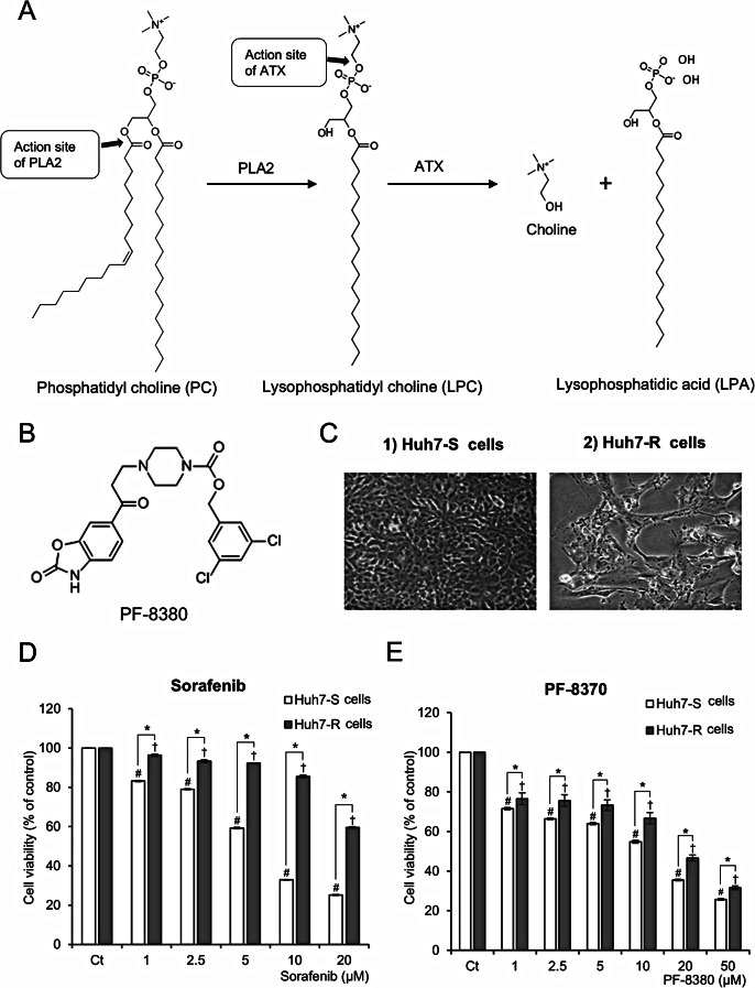
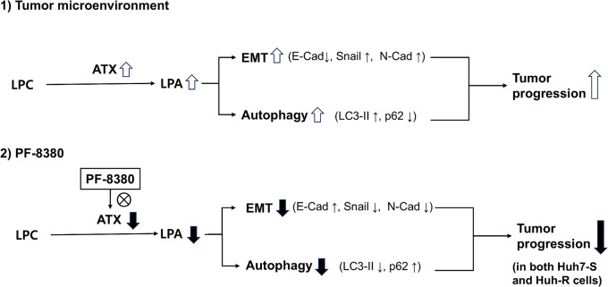
By inhibiting the conversion of lysophosphatidylcholine into lysophosphatidic acid, a process pivotal to tumor progression, the autotaxin (ATX) inhibitor PF-8380 offers a new anticancer therapeutic strategy, distinct from the action mechanism of sorafenib. This study explored the potential anticancer effects of the PF-8380 on hepatocellular carcinoma (HCC) cells, especially sorafenib-resistant strains. The investigation included both in vitro and in vivo experiments to evaluate the impact of PF-8380 treatment on epithelial-mesenchymal transition (EMT) and autophagy markers. An orthotopic HCC model served as the in vivo platform. PF-8380 showed a significant reduction in cell viability in both sorafenib-susceptible and resistant HCC cells. It effectively altered EMT by increasing E-cadherin and reducing Snail levels, and inhibited autophagy, as indicated by changes in LC3 and p62 markers. These effects were consistently observed in the orthotopic HCC mouse model, reinforcing PF-8380's potential as a dual inhibitor of EMT and autophagy in HCC treatment. Our research indicates that PF-8380 could provide substantial therapeutic benefits in the treatment of HCC, even in cases resistant to sorafenib, primarily by suppressing both EMT and autophagy processes.

Keywords: Autophagy; Autotaxin inhibitor; Epithelial-mesenchymal transition (EMT); Hepatocellular carcinoma; Sorafenib.


Conflict of interest statement

Declarations. Ethical approval: The animal studies adhered to the guidelines of the Institute for Laboratory Animal Research, Korea (IRB No: CUMC-2020-0211-06). Competing interests: The authors declare no competing interests.

Figures

Fig. 1
Fig. 1
Antiproliferative effects of PF-8380 in Huh7 cells. (A) Metabolism of phosphatidyl choline (PC), resulting in generation of lysophophatidyl acid (LPA) by autotoxin (ATX). LPA is recognized for facilitating oncogenic processes, including the promotion of cancer cell proliferation, invasive behavior, and metastatic dissemination. (B) Chemical structure of PF-8380, an orally bioavailable piperazinylbenzoxazolone compound acting as an ATX inhibitor. The compound contains two benzene rings, a piperidine ring, an amide group, and a nitrile group. (C) Morphological characteristics of sorafenib-susceptible Huh7 (Huh7-S) cells and sorafenib-resistant Huh7 (Huh7-R) cells during culture. These images depict cellular morphology and do not reflect differences in viability. Cell viability upon PF-8380 treatment was separately quantified in Fig. 1E. (D) Cell viability of Huh7-S and Huh7-R cells after treatment with increasing concentrations of sorafenib (1–20 µM) for 48 h. Huh7-R cells exhibit higher viability than Huh7-S cells, confirming sorafenib resistance. (E) Cell viability of Huh7-S and Huh7-R cells after treatment with increasing concentrations of PF-8380 (1–50 µM) for 48 h. PF-8380 significantly reduces cell viability in a dose-dependent manner in both Huh7-S and Huh7-R cells, indicating its antitumor effect regardless of sorafenib resistance. Values are presented as mean ± SD of three independent experiments. (#P < 0.05) compared to the control (Ct) in Huh7-S cells. (†P < 0.05) compared to the control (Ct) in Huh7-R cells. (*P < 0.05) between Huh7-S and Huh7-R cells at the same treatment concentration
Fig. 2
Fig. 2
Impact of PF-8380 on EMT and cell migration in sorafenib-resistant Huh7 cells. (A) Real-time PCR analysis depicting the alterations in EMT markers post 24-hour treatment with escalating concentrations of PF-8380 (1–10 µM). PF-8380 treatment upregulates the expression of the epithelial marker E-cadherin and downregulates the mesenchymal marker Snail in both Huh7-S and Huh7-R cells. (B) Western blot analysis assessing the changes in EMT markers in response to PF-8380 treatment. A dose-dependent effect is observed with an increase in the expression of the epithelial marker E-cadherin, and a decrease in the expression of the mesenchymal markers N-cadherin and Snail in both cell lines. (C) Cell migration assay showing the inhibitory effect of PF-8380 on the migratory capacity of both Huh7-S and Huh7-R cells, highlighting its potential role in curtailing cancer cell motility and the propensity for metastasis. Data are represented as mean ± SD of three independent experiments. *P < 0.05
Fig. 3
Fig. 3
Autophagy inhibition by PF-8380 in sorafenib-resistant Huh7 cells. (A) Western blot analysis of autophagy markers LC3 and p62 in Huh7-S and Huh7-R cells following treatment with increasing concentrations of PF-8380 (1–10 µM). A trend of decreasing LC3 and increasing p62 levels is observed in correlation with PF-8380 dose escalation. (B) Immunofluorescence of LC3B and p62 in both cell lines pre and post PF-8380 treatment. A marked decrease in LC3B fluorescence and a concurrent increase in p62 is observed post-treatment, indicating an inhibitory effect of PF-8380 on autophagy. Data are represented as mean ± SD of three independent experiments. *P < 0.05
Fig. 4
Fig. 4
Orthotopic HCC mouse models and their response to PF-8380 treatment. (A) Schematic representation of the experimental design. Orthotopic HCC mouse models were established by injecting 5 × 106 Huh7-S and Huh7-R cells into the liver. Three weeks post-tumor implantation, mice were intraperitoneally administered with either 10% DMSO (n = 5) or PF-8380 (10 mg/kg) (n = 5) thrice weekly over a four-week period. (B) Gross morphology of liver specimens collected one week post-treatment. Representative images display the extent of intrahepatic tumor burden in both Huh7-S and Huh7-R mouse models post PF-8380 or DMSO treatment. Notable reduction in tumor cells can be observed in the PF-8380 treated groups
Fig. 5
Fig. 5
PF-8380 attenuates EMT in orthotopic sorafenib-resistant HCC mouse model. (A) Real-time PCR analysis of liver mass specimens revealed that PF-8380 treatment markedly elevated the expression of the epithelial marker, E-cadherin, while concurrently downregulating the expression of the mesenchymal marker, Snail in both Huh7-S and Huh7-R cell-injected cohorts. Data are presented as the mean ± SD, *P < 0.05. (B) Immunohistochemical staining of hepatic tumor tissues further confirmed the upregulation of E-cadherin and downregulation of Snail post PF-8380 administration in both Huh7-S and Huh7-R cell-injected groups. Representative images are shown. Scale bar, 50 μm. Quantification data are shown as the mean ± SD of three independent experiments. *P < 0.05
Fig. 6
Fig. 6
Schematic illustration of the PF-8380-mediated suppression of EMT and autophagy in hepatocellular carcinoma (HCC) cells. (1) In the tumor microenvironment, autotaxin (ATX) catalyzes the conversion of lysophosphatidylcholine (LPC) into lysophosphatidic acid (LPA), leading to the activation of epithelial-mesenchymal transition (EMT) and autophagy. Increased EMT is characterized by decreased E-cadherin (E-Cad) and increased Snail and N-cadherin (N-Cad), while autophagy activation is indicated by increased LC3-II and decreased p62. These processes contribute to tumor progression. (2) PF-8380 inhibits ATX activity, reducing LPA levels, thereby suppressing EMT and autophagy. EMT inhibition is marked by increased E-cadherin and decreased Snail and N-cadherin, while autophagy inhibition is characterized by reduced LC3-II and increased p62. Consequently, tumor progression is suppressed in both sorafenib-sensitive (Huh7-S) and sorafenib-resistant (Huh7-R) HCC cells

References

    1. Balijepalli P, Sitton CC, Meier KE (2021) Lysophosphatidic acid signaling in Cancer cells: what makes LPA so special?? Cells 10. 10.3390/cells10082059 - PMC - PubMed
    1. Bhave SR, Dadey DY, Karvas RM et al (2013) Autotaxin Inhibition with PF-8380 enhances the radiosensitivity of human and murine glioblastoma cell lines. Front Oncol 3:236. 10.3389/fonc.2013.00236 - PMC - PubMed
    1. Chen L, Zhang J, Deng X, Liu Y, Yang X, Wu Q, Yu C (2017) Lysophosphatidic acid directly induces macrophage-derived foam cell formation by blocking the expression of SRBI. Biochem Biophys Res Commun 491:587–594. 10.1016/j.bbrc.2017.07.159 - PubMed
    1. Erstad DJ, Tager AM, Hoshida Y, Fuchs BC (2017) The autotaxin-lysophosphatidic acid pathway emerges as a therapeutic target to prevent liver cancer. Mol Cell Oncol 4:e1311827. 10.1080/23723556.2017.1311827 - PMC - PubMed
    1. Fukushima K, Takahashi K, Yamasaki E, Onishi Y, Fukushima N, Honoki K, Tsujiuchi T (2017) Lysophosphatidic acid signaling via LPA(1) and LPA(3) regulates cellular functions during tumor progression in pancreatic cancer cells. Exp Cell Res 352:139–145. 10.1016/j.yexcr.2017.02.007 - PubMed

MeSH terms