. 2025 Mar 13;16(1):2487.
doi: 10.1038/s41467-025-57566-3.

Epidemiology and transmission dynamics of multidrug-resistant organisms in nursing homes within the United States

Affiliations

Epidemiology and transmission dynamics of multidrug-resistant organisms in nursing homes within the United States

Lona Mody et al. Nat Commun. .

Abstract

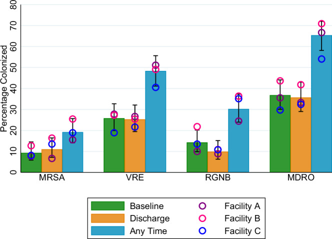
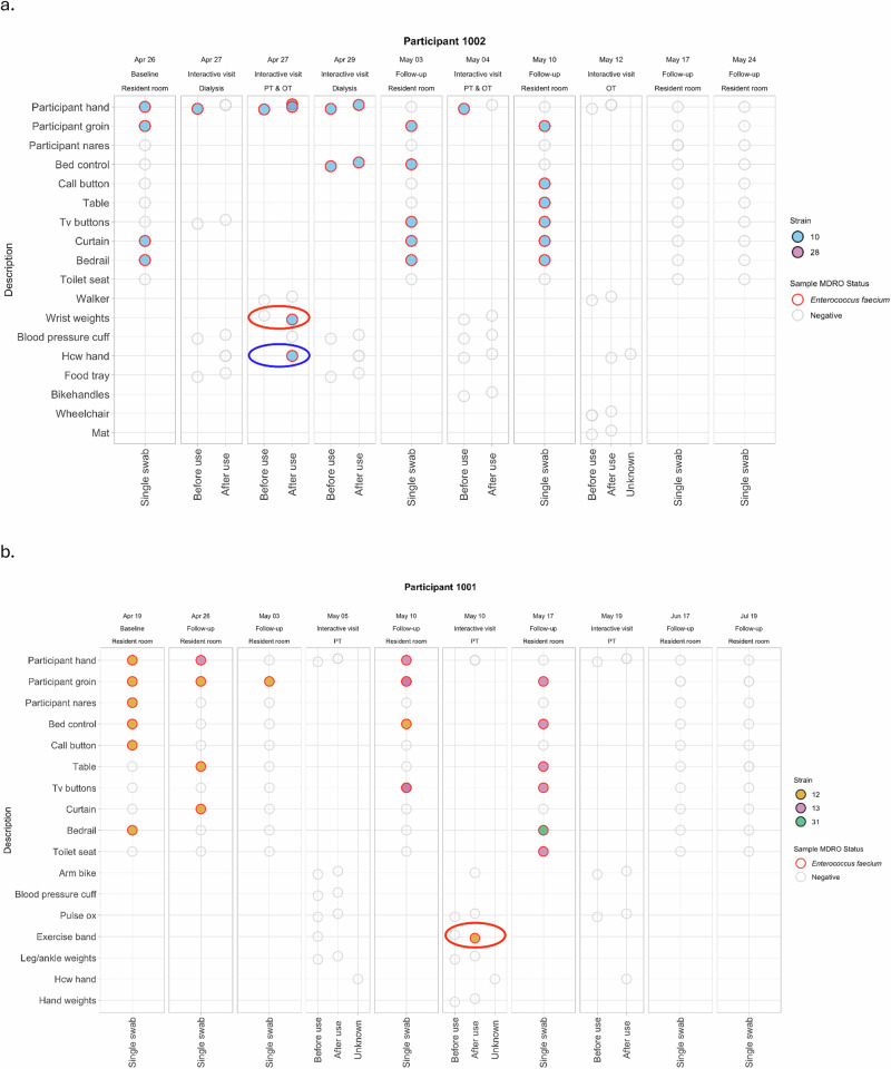
Nursing home (NH) residents in the United States routinely attend interactive visits for services such as therapy or dialysis, creating opportunities for pathogen transmission. A paucity of studies exist which delineate spread of pathogens beyond residents' in-room environment. In this prospective cohort study, we recruited 197 newly-admitted residents across three Veterans Affairs NHs to characterize multidrug-resistant organism (MDRO) prevalence, acquisition, and transmission. Participant hands, nares, groin, and seven environmental surfaces were swabbed during 758 regularly scheduled in-room visits; participant hands, healthcare personnel hands, and equipment were swabbed during 345 unscheduled interactive visits. We demonstrate that baseline MDRO colonization and new acquisition is common, and one in six interactive visits result in MDRO transmission. Whole genome sequencing on a subset of participants enabled us to identify sources of transmission where it was unknown using microbiologic methods alone. Our results illustrate MDRO transmission pathways and highlight the need for innovative, multidisciplinary interventions.


Conflict of interest statement

Competing interests: The authors declare no competing interests.

Figures

Fig. 1
Fig. 1. Colonization with multidrug-resistant organisms at baseline, at discharge, and at any time during the study.
Bars indicate proportion of total participants colonized at baseline (green), discharge (orange), and any time (blue), among participants with >1 in-room visit (n = 182 participants), allowing distinct admission and discharge samples. Dots indicate proportion of participants at Facility A (purple, n = 90 participants), Facility B (pink, n = 55 participants), and Facility C (blue, n = 37 participants) colonized at baseline (green), discharge (orange), and any time (blue), among participants with >1 in-room visit (n = 182 participants). Error bars represent the 95% confidence interval for overall colonization (%), which is the average colonization rate among all three facilities. Based on Pearson’s chi-square tests, no significant differences across the facilities were found. Source data are provided for this figure. Abbreviations: MDRO, multidrug-resistant organism; MRSA, methicillin-resistant Staphylococcus aureus; RGNB, resistant gram-negative bacilli; VRE, vancomycin-resistant enterococci.
Fig. 2
Fig. 2. Changes in MDRO colonization status from baseline visit to discharge (last study visit).
Orange boxes indicate participants colonized with an MDRO, blue boxes indicate participants not colonized with an MDRO. Numbers in each box indicate the number of participants with a given colonization status at study baseline or discharge. For example, 47 participants who had multiple in-room visits were colonized with VRE at baseline. Of those, 27 (57.5%) remained colonized at discharge. The remaining 20 (42.5%) did not have detectable colonization at discharge. Alternatively, 135 participants who had multiple in-room visits were not colonized with VRE at baseline. Of those, 19 (14.1%) acquired VRE during their NH stay, while 116 (85.9%) remained not colonized with VRE. Source data are provided for this figure. Abbreviations: MDRO, multidrug-resistant organism; MRSA, methicillin-resistant Staphylococcus aureus; NH, nursing home; RGNB, resistant gram-negative bacilli; VRE, vancomycin-resistant enterococci.
Fig. 3
Fig. 3. Transmission of DNA-confirmed MRSA or VRE to participant hands or equipment.
a MRSA transmission to equipment. Of the 14 interactive visits where the participant hand was contaminated with MRSA before use, transmission of MRSA to a surface occurred at 4 (28.6%) visits. Ten MRSA transmissions to a surface occurred, of which 4 (40%) can be attributed to participant hand contamination. b MRSA transmission to participant hand. Of the 17 interactive visits where equipment or surface was contaminated with MRSA before use, transmission of MRSA to the participant’s hand occurred at 1 (5.9%) visit. Ten MRSA transmissions to a participant hand occurred, of which 1 (10%) can be attributed to surface contamination. c VRE transmission to equipment. Of the 36 interactive visits where the participant hand was contaminated with VRE before use, transmission of VRE to a surface occurred at 16 (44.4%) visits. Twenty-five VRE transmissions to a surface occurred, of which 16 (64.0%) can be attributed to participant hand contamination. d VRE transmission to participant hand. Of the 35 interactive visits where equipment or surface was contaminated with VRE before use, transmission of VRE to the participant’s hand occurred at 2 (5.7%) visits. Thirteen VRE transmissions to a participant hand occurred, of which 2 (15.4%) can be attributed to surface contamination. e Unadjusted odds ratios and confidence intervals for the association between surface contamination and participant hand colonization and transmission of MRSA and VRE. Reported p-values (two-sided) are based on generalized linear models specifying an exchangeable, within-participant correlation structure for the panels. Transmission of MRSA to a surface/equipment was 20.7 times more likely when the participant hand was colonized first, compared to when the participant hand is not colonized first (p = 2.5e-5), while transmission of MRSA to a participant hand is 2.2 times more likely when the surface/equipment is contaminated first (p = 0.451). Transmission of VRE to a surface/equipment was 21.3 times more likely when the participant hand was colonized first, compared to when the participant hand is not colonized first (p = 4.1e−10), while transmission of VRE to a participant hand is 1.7 times more likely when the surface/equipment is contaminated first (p = 0 .505). Source data are provided for this figure. Abbreviations: MRSA, methicillin-resistant Staphylococcus aureus; VRE, vancomycin-resistant enterococci; OR, odds ratio.
Fig. 4
Fig. 4. Genomics support of microbiological linkages among two participants.
Column headings indicate date, location, and type (baseline or follow-up = in-room; or interactive) of each study visit. Transmission events are identified with a red circle; single positive swabs not able to be assessed for transmission are identified with a blue circle. White circles outlined in gray are samples collected and negative for any MDRO; colored circles outlined in red are samples collected and positive for a particular strain (listed in each legend). a For participant 1002, VRE strain 10 was detected at multiple body sites (groin and hand) and environmental surfaces (bedrail and privacy curtain) at study baseline (Apr 26). Study staff attended a dialysis session with this participant on Apr 27, during which time the participant’s hand was colonized with VRE strain 10 at the start of the session, but no transmission of VRE to any surfaces was detected during that interactive visit. Study staff also attended a PT & OT combined session on that same day (Apr 27); during this interactive visit, a VRE strain 10 transmission (circled in red) was detected at the wrist weights, since the weight went from VRE-negative to VRE-positive following participant use. Based on our microbiology results, the participant’s hand is the likely source of this transmission, since the participant’s had was VRE-positive at the beginning of the interactive visit. Furthermore, sequencing confirms these VRE strains are identical. The healthcare provider hand at the end of the session was also VRE-positive; however, this is not counted as a transmission because we do not have a “before” swab on the healthcare provider hand to prove that it changed status from negative to positive. Sequencing results confirm this strain is identical to that carried on the participant hand, highly suggestive of transmission. This participant continued to be colonized, and their in-room surface contaminated, at two subsequent in-room visits. This participant’s hand was also colonized during two subsequent interactive visits, but no other transmissions were detected. b For participant 1001, VRE strain 12 was detected at multiple body sites (nares, groin, and hand) and environmental surfaces (bed control, bedrail, call button) at study baseline (on Apr 19). During the first in-room follow-up visit (Apr 26), VRE strain 12 was detected at the participant groin, table top, and privacy curtain, while VRE strain 13 was detected on the participant’s hand. During the next in-room follow-up visit (May 3), VRE strain 12 was detected only at the participant groin. Study staff next attended a PT session with this participant (May 5), during which time no MDROs were found on the participant’s hand, on surfaces, nor on the healthcare provider hand. The next visit with this participant was in his or her room (May 10), where VRE strain 13 was detected at the participant’s groin, hand, and TV remote, while VRE strain 12 was detected at the bed control. Study staff attend a PT session with this participant next (on May 10), during which time a VRE strain 12 transmission (circled in red) was detected at the exercise band, since the exercise band changed status from VRE-negative to VRE-positive following participant use. The participant’s hand was not positive for VRE at the start of this session, so we cannot assume the source of this transmission was the participant’s hand. However, sequencing allows us to speculate that the participant’s hand is the likely source, since their hand was colonized with VRE strain 12 at an earlier, in-room visit (on Apr 19). This participant continued to be colonized (VRE strain 13), and their in-room surface contaminated (VRE strains 13 and 31), at one subsequent in-room visit. This participant’s hand was not colonized at any subsequent interactive visits. Abbreviations: MDRO, multidrug-resistant organism; OT, occupational therapy; PT, physical therapy; VRE, vancomycin-resistant enterococci.
Fig. 5
Fig. 5. A representative interactive visit that resulted in a transmission event.
This participant, who was colonized with VRE at the hand, visited the rehabilitation gym for physical and occupational therapy. The first piece of equipment used by the participant was a hand weight; the hand weight was negative for any MDRO prior to use and was contaminated with VRE after approximately 6 minutes of participant contact. This change in surface contamination is an example of a transmission event, with the source being the participant’s hand and the destination surface being the weight. The participant’s hand remained colonized with VRE during the session. The walker was used next by the participant -- it was negative for any MDRO prior to use and after two minutes of use, no transmission occurred. The last item of contact, the blood pressure cuff, was not swabbed before use, but was negative for any MDRO after being on the participant’s arm for five minutes. The bare hand of the physical therapist was negative for any MDRO after taking the participant’s blood pressure. The occupational therapist’s gloved hand was positive for VRE at the end of the session, following several touches to the participant and walker. Because we only swabbed the occupational therapist’s hand one time, we cannot conclusively say a transmission occurred (as we do not know if their hand was negative at the session start). The participant’s hand remained colonized with VRE at the end of the session. “Nurse” icon by Llisole from https://thenounproject.com/browse/icons/term/nurse/ CC BY 3.0. All other icons downloaded from thenounproject.com via a paid subscription (no attribution required).

References

    1. Werner, R. M., Templeton, Z., Apathy, N., Skira, M. M. & Konetzka, R. T. Trends in post-acute care in U.S. nursing homes: 2001-2017. J. Am. Med Dir. Assoc.22, 2491–2495 (2021). - PMC - PubMed
    1. Werner, R. M. & Bressman, E. Trends in post-acute care utilization during the COVID-19 pandemic. J. Am. Med Dir. Assoc.22, 2496–2499 (2021). - PMC - PubMed
    1. White, H. K. Post-acute care: Current state and future directions. J. Am. Med Cir. Assoc.20, 392–395 (2019). - PubMed
    1. Mody, L., Bradley, S. F. & Huang, S. S. Keeping the “home” in nursing home: Implications for infection prevention. JAMA Intern Med173, 853–854 (2013). - PMC - PubMed
    1. McKinnell, J. A. et al. High prevalence of multidrug-resistant organism colonization in 28 nursing homes: An “Iceberg Effect. J. Am. Med Dir. Assoc.21, 1937–1943 (2020). - PMC - PubMed

Substances