. 2025 Apr;17(4):696-721.
doi: 10.1038/s44321-025-00208-4. Epub 2025 Mar 13.

Immune training enhances anti-viral responses and improves outcomes in Pax5-/+ mice susceptible to chronic infection

Affiliations

Immune training enhances anti-viral responses and improves outcomes in Pax5-/+ mice susceptible to chronic infection

Zhe Lu et al. EMBO Mol Med. 2025 Apr.

Abstract

Viral infections pose a significant global burden. Host susceptibility to pathogens is determined by many factors including genetic variation that can lead to immunodeficient or dysregulated antiviral immune responses. Pax5 heterozygosity (Pax5-/+), resulting in reduced PAX5 levels in mice, mimics germline or somatic PAX5 dysregulation contributing to diseases such as childhood B-cell precursor acute lymphoblastic leukemia (B-ALL). In contrast to the well-characterized roles of PAX5 during early B-cell development, little is known about how Pax5 heterozygosity impacts antiviral responses. We infected Pax5-/+ mice with the noncytopathic Lymphocytic Choriomeningitis Virus (LCMV) and found that infection with the chronic Docile strain resulted in decreased survival of Pax5-/+ mice. While early adaptive CD8+ T-cell (CTL) immunity was robust in Pax5-/+ mice, LCMV-specific neutralizing antibody production was compromised leading to impaired long-term viral clearance and a pro-inflammatory milieu in the bone marrow (BM). Here we show that survival outcomes were improved upon prophylactic treatment with the β-glucan immune trainer through induction of heterologous protection against chronic infection. β-Glucan enhanced viral clearance, CTL immunity, neutralizing antibody production and reduced monocyte immunosuppression in multiple LCMV-resident host organs. New insight from this study will help design effective prophylactic treatment strategies against chronic viral infections, particularly in genetically predisposed susceptible hosts.

Keywords: Chronic Infection; LCMV; PAX5; Trained Immunity; β-glucan.


Conflict of interest statement

Disclosure and competing interests statement. PAL, KSL, HCX, and PP declare that they are involved in the development of LCMV for clinical application in oncology in cooperation with, as founders of, or as advisors to Abalos Therapeutics GmbH. The remaining authors declare no competing interests.

Figures

Figure 1
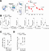
Figure 1. Pax5 haploinsufficiency confers an increased susceptibility to chronic but not acute viral infection.
(A) Percentages of naive CD4+ and CD8+ T cells, memory and activated T cells, and NK cells in PBMCs of three healthy carriers and one patient (with PAX5 loss of heterozygosity (LOH) in remission more than 5 years after completion of therapy) compared to reference healthy control ranges (gray box) are shown. (B) Pax5−/+ and WT mice were intravenously infected with 106 PFU of VSV and survival was monitored (n ≥10 mice per group). (CE) Pax5−/+ and WT mice were intravenously infected with 106 PFU of LCMV Docile and (C) survival was monitored (n ≥12 mice per group) *P = 0.0002, #P = 0.0384. Statistical analyses were performed using Log-rank (Mantel–Cox) test with a Bonferroni correction for multiple curve comparisons. Where material was available, virus titers in the bone marrow (BM) and spleen (D) as well as LDH levels in the plasma (E, n = 6) were determined from mice in the survival curve in (C). S indicates that the mice were necessarily sacrificed as they succumbed to infection (n ≥9). C indicates healthy mice which were sacrificed as accompanying controls and/or at endpoint (n ≥13). Unless otherwise stated, statistical analyses were performed using a Student´s t test (unpaired, two-tailed). Error bars indicate SEM. Source data are available online for this figure.
Figure 2
Figure 2. Pax5 heterozygosity does not impair initial antiviral CTL responses but impacts long-term virus control.
(AE) Pax5−/+ and WT mice were intravenously infected with 106 PFU of LCMV Docile and (A) Numbers of effector CTL tet-gp33+ and tet-np396+ CD8 T cells were measured at day 15 post virus inoculation in the liver, spleen lymph node (LN), and bone marrow (BM) using FACS (n ≥3 mice per group). (B) Effector tet-gp33+ and tet-np396+ CD8 T cells were measured in the blood at the indicated days post infection (n ≥3 mice per group). (C) LCMV virus titers were determined in the spleen, bone marrow (BM), lymph nodes (LN), and liver tissue at day 15 (n = 4) and 120 post infection using the plaque assay (n = 11). (D) LDH activity in the plasma was determined at day 15 (n = 4) and day 120 post infection (n ≥ 6). (E, left panel) Sections of snap-frozen liver tissues were analyzed using H&E staining (a representative set of images of n ≥3 mice per group is shown; scale bar = 50 μm; arrows indicate the inflammatory infiltrates). (E, right panel) Infiltrates were quantified. Error bars indicate SEM. Unless otherwise stated, all statistical analyses were performed by a Student´s t test (unpaired, two-tailed). Source data are available online for this figure.
Figure 3
Figure 3. Pax5 haploinsufficiency leads to impaired anti-LCMV neutralizing antibody production during chronic infection.
(A) B-cell gating strategy for the assessment of mature splenic B cells is shown. Numbers of B1, marginal zone (MZ), follicular (FO), and transitional (T1, T2, and T3) B cells were assessed in the spleen of naive WT and Pax5-/+ mice (n = 4 mice per group). (BD) Immunoglobulins were evaluated at the indicated time points using the 6-PLEX Mouse Immunoglobulin Isotyping Panel (n = ≥3 mice per group). (C) LCMV-neutralizing antibodies in the plasma were indirectly assessed using plaque assay on the indicated days (n ≥4 mice per group). (D) LCMV-specific neutralizing antibodies against the LCMV glycoprotein (GP) were assessed using ELISA on the indicated days, showing the fold increase from the background (naive serum) on the indicated days (n = 4 mice per group). Error bars indicate SEM. All statistical analyses were performed using a Student´s t test (unpaired, two-tailed). Source data are available online for this figure.
Figure 4
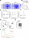
Figure 4. Pax5 haploinsufficiency shapes a distinct bone marrow microenvironment (BME) in response to chronic infection.
(A) Cytokine levels in the bone marrow and plasma were evaluated using the Procarta 19-Plex assay in uninfected naive controls (day 0), day 15 and day 120 post infection with 106 PFU of LCMV Docile. Fold changes relative to day 0 genotype-specific controls are represented in a heatmap (n = 3 mice per group). *As indicated in the figure represents significant differences relative to time point 0 within each genotype as determined by a one-way ANOVA with a Dunnett’s post hoc test. IL-18 (day 15 P = <0.0001, day 120 P = 0.0471), CXCL10 (P = 0.0297), CXCL9 (P = 0.0467), CCL3 (P = 0.0003), CCL4 (day 15 BM WT P = 0.0016, day 15 plasma WT P < 0.0001, day 120 plasma Pax5-/+ (P = 0.0401), CCL5 (P = 0.0016), CXCL1 (BM P = 0.0079, plasma day 15 P = 0.0002, plasma day 120 P = 0.0033), IFNγ (Pax5−/+ P = 0.0344, WT P = 0.0346). #As indicated in the figure represents significant differences between the Pax5−/+ and WT group at the indicated time point as determined by a Student´s t test (unpaired, two-tailed). IL-18 (P = 0.038), CXCL10 (P = 0.009), CXCL9 (P = 0.006), CCL3 (BM P = 0.017, plasma P = 0.004), CCL4 (P = 0.020), CXCL1 (P = 0.01), IFNγ (P = 0.037). (B) Pax5−/+ and WT mice were intravenously infected with 106 pfu of LCMV Docile and effector CTL tet-gp33+ and tet-np396+ CD8 T cells were measured in the bone marrow at the indicated days post infection (n ≥3 mice per group). (C) Surface molecule PD1, LAG3 and TIM-3 expression was measured on tet+ CD8 T cells in the bone marrow at the indicated time points (n ≥ 3 mice per group). (D) B220+CD138+ and B220-CD138+ cells were evaluated in the bone marrow as was the percent distribution of CD28 within the B220-CD138+ cell population 120 days post infection (n ≥ 6 mice per group). (E) B220-CD138+ cells from (D) were further subdivided into percentages of CD19 and CD19+ cells (n ≥ 2 mice per group). Error bars indicate SEM. Unless otherwise stated, all statistical analyses were performed using a Student´s t test (unpaired, two-tailed). (F) The effects of chronic infection in Pax5+/− and WT mice are summarized (created in BioRender. Mescher M (2025) https://BioRender.com/b91v085). Source data are available online for this figure.
Figure 5
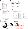
Figure 5. Immune training with β-glucan improves outcomes following chronic LCMV infection.
(A) WT and Pax5−/+ mice were injected with 1 mg of β-glucan and LSK (Lin-Sca-1+c-KIT+) cells were measured in the bone marrow 24 h post treatment (n = 3 mice per group). (B) A schematic representation of the treatment regimen is shown (Created in BioRender. Mescher M (2025) https://BioRender.com/v92i052). (C) Pax5+/− and WT mice were injected with 1 mg of β-glucan. Seven days later, this was followed by infection with 106 PFU of LCMV Docile and survival was monitored (n ≥ 6 mice per group). Statistical analyses were performed using a Log-rank (Mantel–Cox) test with a Bonferroni correction for comparisons. (D) Virus titers were determined in the bone marrow (BM), spleen and lymph nodes (LN) of Pax5-/+, and WT mice from the survival curve in (C). S indicates that the mice were sacrificed before the endpoint and succumbed to infection (n = 3 mice). C indicates healthy mice which were sacrificed as accompanying controls and at endpoint (n ≥ 6 mice). (E) Survival proportions of LCMV Docile Pax5-/+ and WT mice from Fig. 1B are directly compared to β-glucan pre-treated LCMV Docile-infected mice in (C). Error bars indicate SEM. Unless otherwise stated, all statistical analyses were performed using a Student´s t test (unpaired, two-tailed). Source data are available online for this figure.
Figure 6
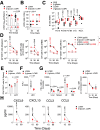
Figure 6. Immune training with β-glucan improves T-cell immunity and affects immunosuppressive infiltrates following chronic LCMV infection.
(BF) WT mice were injected with 1 mg of β-glucan and 7 days later this was followed by infection with 106 PFU of LCMV Docile. (A) LCMV virus titers were determined in the spleen at day 15 (n = 3 mice per group) and day 30 post LCMV infection using plaque assay (n ≥ 7 mice per group). (B) LCMV-neutralizing antibodies in the plasma were indirectly assessed using plaque assay on day 30 (n = 7 mice per group) and 60 post infection (n ≥ 3 mice per group). (C) Pro-B, Pre-BI, Pre-BII, immature, and re-circulating B cells were measured in the bone marrow at day 15 post infection (n ≥ 3 mice per group). Statistical analyses were performed using a one-way ANOVA with a Tukey post hoc test. (D) Numbers of effector CTL tet-gp33+ and tet-np396+ CD8 T cells were measured using FACS at the indicated time points in the blood, spleen, liver, and bone marrow (n ≥ 3 mice per group). (E) 15 days post infection, single cell suspended splenic cells were restimulated with the gp33 LCMV-specific epitope, followed by intracellular staining for IFN-γ (n = 3 mice per group). Statistical analysis was performed using a one-way ANOVA with a Dunnett’s post hoc test. (F) LDH activity in the plasma was determined 15 days post infection (n ≥ 4 mice per group). (G) PD-L1 expression was determined on monocytes (CD11B+Ly6GlowLy6Chigh) in the indicated organs (n = 4 mice per group) and blood (n ≥ 3 mice per group) 30 days post infection. (H) Cytokine and chemokine levels in the plasma were evaluated using the Procarta 19-Plex assay in at day 15 post infection and significant differences between the β-glucan pre-treated infected and the infected alone group are shown (n ≥ 5 mice per group). Error bars indicate SEM. Unless otherwise stated, all statistical analyses were performed using a Student´s t test (unpaired, two-tailed). Source data are available online for this figure.
Figure 7
Figure 7. β-glucan maintains some protective effects against chronic LCMV when administered post infection.
(A) WT mice were infected with 106 PFU of LCMV Docile and 7 days later treated with 1 mg of β-glucan. Created in BioRender. Mescher M (2025) https://BioRender.com/p95y241 (B) LDH activity in the plasma was determined 15 days post infection (n = 6 mice per group). (C) LCMV virus titers were determined in the spleen and liver at day 30 post LCMV infection using plaque assay (n ≥ 5 mice per group). (D) Number of effector CTL tet-gp33+ T cells were determined in the spleen using FACS analysis 30 days post infection (n = 5–6). (E) LCMV-neutralizing antibodies in the plasma were indirectly assessed using plaque assay on day 30 post infection (n = 6 mice per group). (FH) WT mice were injected with 1 mg of β-glucan, and 7 days later this was followed by infection with 104 PFU of LCMV Docile. One infected-only group was also treated with 1 mg of β-glucan 7 days post infection. (F) PD-L1 expression was determined on monocytes in the indicated organs 30 days post infection (n = 5 mice per group). Statistical analyses were performed using a one-way ANOVA with a Tukey post hoc test. (G) 30 days post infection, singly suspended cells from the lymph node (LN) or liver were restimulated with the gp33 LCMV-specific epitope, followed by intracellular staining for granzyme B expression (GZMB) (n ≥ 4 mice per group). Statistical analyses (F, G) were performed using a one-way ANOVA with a Tukey post hoc test. (H) Expression of KLRG1 on CD8+ T cells in the spleen, blood, and liver was assessed 30 days post infection (n = 5 mice per group). (I) Effector CTL tet-gp33+ T cells (expressed as percent of CD8+ T cells) were assessed in the spleen, blood and liver 30 days post infection (n = 5). Statistical analyses (H, I) were performed using a one-way ANOVA with a Dunnett’s post hoc test (J) Splenic sections were stained with the indicated antibodies and detected using CO-Detection by indEXing (CODEX), scale bar indicates 50 μm; (a representative image of n of 5 (mice per group) is shown). Error bars indicate SEM. Unless otherwise stated, statistical analyses were performed using a Student´s t test (unpaired, two-tailed). Source data are available online for this figure.
Figure EV1
Figure EV1. Pax5 haploinsufficiency does not compromise hosts following VSV and acute LCMV infection.
(A) WT and Pax5−/+ mice were infected with 106 PFU of VSV and IFN-α concentration was determined in the plasma of WT (n = 3 mice per group) and Pax5+/− mice (n = 4 mice per group) 24 h after infection. WT and Pax5−/+ mice were infected with 106 PFU of LCMV-Armstrong strain (Arm.) and (B) viral titers were determined 15 days post infection using the plaque assay (n = 3 mice per group). Statistical analyses were performed using a Student´s t test (unpaired, two-tailed). (C) Survival was monitored (n = 3 mice per group). Statistical analysis was performed a Log-rank (Mantel–Cox) test with a Bonferroni correction for comparisons. (D) FACS blots of spleen, bone marrow and lymph node tissue showing the T-ALL that developed at day 182 post LCMV infection in a Pax5−/+ mouse. Error bars indicate SEM. Source data are available online for this figure.
Figure EV2
Figure EV2. Pax5 haploinsufficiency shapes distinct cytokine profiles in the plasma and bone marrow microenvironment in response to chronic infection.
Cytokine levels in the bone marrow and plasma were evaluated using the Procarta 19-Plex assay in uninfected naive controls (day 0), day 15 and day 120 post infection with 106 PFU of LCMV Docile in Pax5−/+ and WT mice (n = 3 mice per group). Concentrations in pg/ml of detectible cytokines are shown. *As indicated in the figure represents significant differences relative to time point 0 within each genotype as determined by a one-way ANOVA with Dunnett’s post hoc test. IFNγ (BM P = 0.0036, plasma P = 0.045), IL-18 (P < 0.0001), CXCL10 (P = 0.0002), CCL3 (P = 0.004), CCL5 (BM day 15 P = 0.0105, day 120 P = 0.0155, plasma P < 0.0001), IL-12 (P = 0.0018), CXCL1 (BM P = 0.0358, plasma P = 0.0081), CCL4 (BM P = 0.0150, plasma P < 0.0001). #As indicated in the figure represents significant differences between the Pax5−/+ and WT group at a given time point as determined by a Student´s t test (unpaired, two-tailed). CXCL10 (P = 0.003), CCL5 (P = 0.030), IL-12 (P = 0.028). Error bars indicate SEM. Source data are available online for this figure.
Figure EV3
Figure EV3. Chronic LCMV infection affects early B cells populations in the bone marrow of Pax5-/+ and WT hosts.
Pax5−/+ and WT mice were intravenously infected with 106 PFU of LCMV Docile. (A) Pro-B, Pre-BI and Pre-BII cells were measured in the bone marrow at the indicated days post infection using FACS (n ≥ 3 mice per group). (Pro-B, WT P = 0.0007 Pax5−/+ P = 0.0010), (Pre-BI, WT P = 0.001 Pax5−/+ P = 0.0007), (Pre-BII, WT P = 0.0055 Pax5−/+ P = 0.0037). (B) Surface molecule MHC-II and IL-7r expression was measured on Pro-B, Pre-BI and Pre-BII cells in the bone marrow 90 days post infection (n ≥ 3 mice per group). Statistical analyses for (A, B) were performed using a one-way ANOVA with a Tukey post hoc test. (C) PAX5 expression data in different human B-cell subsets was mined from The Human Protein Atlas, Monaco dataset (n = 4). (D) B220+CD138+ and B220-CD138+ numbers were evaluated in the bone marrow of WT (n = 7 mice per group) and Pax5−/+ mice (n = 6) 120 days post infection using FACS analysis. Error bars indicate SEM. Statistical analysis was carried out using a Student´s t test (unpaired, two-tailed). Source data are available online for this figure.
Figure EV4
Figure EV4. Immune training with β-glucan improves T-cell immunity following chronic LCMV infection.
(A) WT mice were injected with 1 mg of β-glucan and LSK (Lin-Sca-1+c-KIT+) cells were measured in the bone marrow 24 h and 15 days post treatment (n ≥ 3 mice per group). (B) WT mice were injected with 1 mg of β-glucan and 7 days later this was followed by infection with 106 PFU of LCMV Docile. LCMV virus titers were determined in the kidney, lymph node (LN) and liver at day 15 (n = 3 mice per group) and 30 (n ≥ 7 mice per group) days post LCMV infection using plaque assay. Statistical analysis of the contingency table was done using a Fisher’s exact test. (C) Numbers of B1, marginal zone (MZ), follicular (FO) and transitional (T1, T2 and T3) B cells were assessed in naive (n = 4 mice per group) and β-glucan (n = 5 mice per group) treated mice at day 30 post β-glucan treatment. (D) Splenic sections were stained with the indicated antibodies and detected using CO-Detection by indEXing (CODEX), scale bar indicates 100 μm; (a representative image of n of 5 (mice per group) is shown). Error bars indicate SEM. Unless otherwise stated, statistical analyses were performed using a Student´s t test (unpaired, two-tailed). Source data are available online for this figure.
Figure EV5
Figure EV5. Immune training with β-glucan affects immunosuppressive infiltrates following chronic LCMV infection.
WT mice were injected with 1 mg of β-glucan and 7 days later this was followed by infection with 106 PFU of LCMV Docile. (A) 30 days post infection, numbers of Treg’s (CD4+CD25+) as well as (B) Treg FOXP3 expression were measured using FACS analysis in the spleen, bone marrow (BM) and lymph node (LN) of infected mice (n ≥ 6 mice per group). (C) 30 Days post infection, frequencies of monocytes (CD11B+Ly6GlowLy6Chigh) were measured using FACS analysis in the spleen, bone marrow (BM) and lymph node (LN), liver (n = 4 mice per group) and in the blood at the indicated time points (n ≥ 3 mice per group). Error bars indicate SEM. Error bars indicate SEM. Statistical analyses were performed using a Student´s t test (unpaired, two-tailed). Source data are available online for this figure.

References

    1. Ali MF, Driscoll CB, Walters PR, Limper AH, Carmona EM (2015) Beta-glucan-activated human B lymphocytes participate in innate immune responses by releasing proinflammatory cytokines and stimulating neutrophil chemotaxis. J Immunol 195:5318–5326 - PMC - PubMed
    1. Auer F, Ruschendorf F, Gombert M, Husemann P, Ginzel S, Izraeli S, Harit M, Weintraub M, Weinstein OY, Lerer I et al (2014) Inherited susceptibility to pre B-ALL caused by germline transmission of PAX5 c.547G>A. Leukemia 28:1136–1138 - PubMed
    1. Auinger A, Riede L, Bothe G, Busch R, Gruenwald J (2013) Yeast (1,3)-(1,6)-beta-glucan helps to maintain the body’s defence against pathogens: a double-blind, randomized, placebo-controlled, multicentric study in healthy subjects. Eur J Nutr 52:1913–1918 - PMC - PubMed
    1. Calderon L, Schindler K, Malin SG, Schebesta A, Sun Q, Schwickert T, Alberti C, Fischer M, Jaritz M, Tagoh H et al (2021) Pax5 regulates B cell immunity by promoting PI3K signaling via PTEN down-regulation. Sci Immunol 6:eabg5003 - PMC - PubMed
    1. Cane S, Ugel S, Trovato R, Marigo I, De Sanctis F, Sartoris S, Bronte V (2019) The endless saga of monocyte diversity. Front Immunol 10:1786 - PMC - PubMed

MeSH terms

Substances

LinkOut - more resources