

Human BioMolecular Atlas Program (HuBMAP): 3D Human Reference Atlas construction and usage
- PMID: 40082611
- PMCID: PMC11978508
- DOI: 10.1038/s41592-024-02563-5
Human BioMolecular Atlas Program (HuBMAP): 3D Human Reference Atlas construction and usage
Abstract
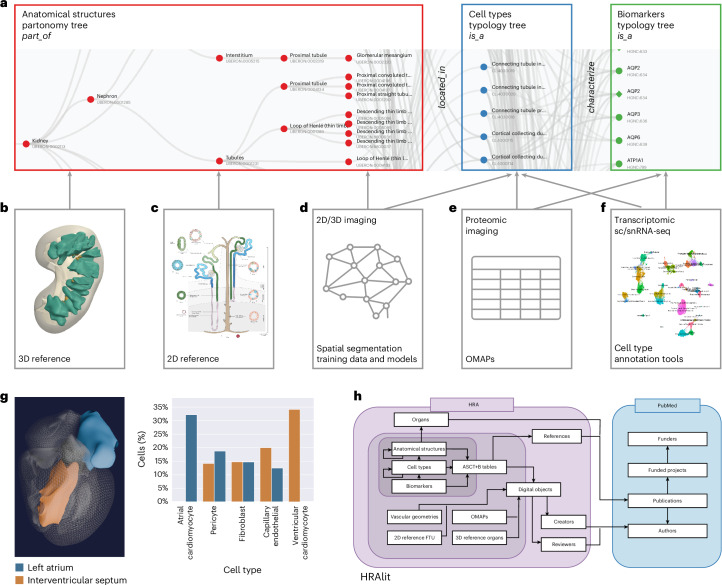
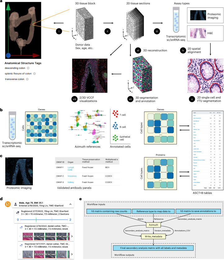
The Human BioMolecular Atlas Program (HuBMAP) aims to construct a 3D Human Reference Atlas (HRA) of the healthy adult body. Experts from 20+ consortia collaborate to develop a Common Coordinate Framework (CCF), knowledge graphs and tools that describe the multiscale structure of the human body (from organs and tissues down to cells, genes and biomarkers) and to use the HRA to characterize changes that occur with aging, disease and other perturbations. HRA v.2.0 covers 4,499 unique anatomical structures, 1,195 cell types and 2,089 biomarkers (such as genes, proteins and lipids) from 33 ASCT+B tables and 65 3D Reference Objects linked to ontologies. New experimental data can be mapped into the HRA using (1) cell type annotation tools (for example, Azimuth), (2) validated antibody panels or (3) by registering tissue data spatially. This paper describes HRA user stories, terminology, data formats, ontology validation, unified analysis workflows, user interfaces, instructional materials, application programming interfaces, flexible hybrid cloud infrastructure and previews atlas usage applications.
© 2025. The Author(s).
Conflict of interest statement
Competing interests: The primary authors declare the following competing interests: R. Satija receives compensation from 10x Genomics, Parse Biosciences and Neptune Bio. R.S. is a co-founder and equity holder of Neptune Bio. S. Teichmann is a remunerated member of the Scientific Advisory Boards of QIAGEN, Foresite Labs and Element Biosciences, a co-founder and equity holder of TransitionBio and EnsoCell Therapeutics and a part-time employee of GlaxoSmithKline since January 2024. The HRA Team authors declare the following competing interests: B. Aronow declares Nexstone Immunology, Uniquity and Advisors. C. Werlein declares speaker fees from Boehringer Ingelheim. M. Snyder declares Personalis, SensOmics, Qbio, January AI, Fodsel, Filtricine, Protos, RTHM, Iollo, Marble Therapeutics, Crosshair Therapeutics, NextThought and Mirvie, Jupiter, Neuvivo, Swaza, Mitrix, Yuvan, TranscribeGlass and Applied Cognition. N. Kelleher declares Thermo Fisher Scientific, Proteinaceous, Integrated Protein Technologies and ImmPro. W. Müller declares Miltenyi Biotec. E. Lundberg is an advisor for the Chan-Zuckerberg Initiative Foundation, Element Biosciences, Cartography Biosciences, Pfizer and Pixelgen Technologies. T. Kendall serves as a consultant or advisory board member for Resolution Therapeutics, Clinnovate Health, HistoIndex, Fibrofind, Kynos Therapeutics, Perspectum, Concept Life Sciences and Jazz Pharmaceuticals; and has received speakers' fees from Servier Laboratories, Jazz Pharmaceuticals, Astrazeneca, HistoIndex and Incyte Corporation. A. Ropelewski is an equity holder in Illumina, Nanostring, 10x Genomics and Akoya. L. Falo is a cofounder and equity holder in SkinJect. The remaining authors declare no competing interests.
Figures
Update of
-
Human BioMolecular Atlas Program (HuBMAP): 3D Human Reference Atlas Construction and Usage.bioRxiv [Preprint]. 2024 Aug 14:2024.03.27.587041. doi: 10.1101/2024.03.27.587041. bioRxiv. 2024. Update in: Nat Methods. 2025 Apr;22(4):845-860. doi: 10.1038/s41592-024-02563-5. PMID: 38826261 Free PMC article. Updated. Preprint.
References
MeSH terms
Substances
Grants and funding
- U54 HL165443/HL/NHLBI NIH HHS/United States
- OT2 OD033759/OD/NIH HHS/United States
- U54 AG075936/AG/NIA NIH HHS/United States
- OT2 OD033761/OD/NIH HHS/United States
- RM1HG011014/U.S. Department of Health & Human Services | NIH | National Human Genome Research Institute (NHGRI)
- U54 AG075932/AG/NIA NIH HHS/United States
- U24CA268108/U.S. Department of Health & Human Services | National Institutes of Health (NIH)
- U01 HL148861/HL/NHLBI NIH HHS/United States
- OT2 OD026675/OD/NIH HHS/United States
- OT2 OD033760/OD/NIH HHS/United States
- OT2OD033760/U.S. Department of Health & Human Services | National Institutes of Health (NIH)
- R03 OD036499/OD/NIH HHS/United States
- U24 DK135157/DK/NIDDK NIH HHS/United States
- U54HL165443/U.S. Department of Health & Human Services | National Institutes of Health (NIH)
- OT2OD033759/U.S. Department of Health & Human Services | National Institutes of Health (NIH)
- OT2OD026671/U.S. Department of Health & Human Services | National Institutes of Health (NIH)
- RM1 HG011014/HG/NHGRI NIH HHS/United States
- U2C DK114886/DK/NIDDK NIH HHS/United States
- OT2 OD026671/OD/NIH HHS/United States
- U01 DK133090/DK/NIDDK NIH HHS/United States
- OT2OD026673/U.S. Department of Health & Human Services | National Institutes of Health (NIH)
- OT2 OD026682/OD/NIH HHS/United States
- U54 AR081775/AR/NIAMS NIH HHS/United States
- OT2 OD033756/OD/NIH HHS/United States
- 3U54AG075936/U.S. Department of Health & Human Services | National Institutes of Health (NIH)
- 3OT2OD026682/U.S. Department of Health & Human Services | National Institutes of Health (NIH)
- U24DK135157/U.S. Department of Health & Human Services | National Institutes of Health (NIH)
- OT2 OD026673/OD/NIH HHS/United States
- 1R03OD036499/U.S. Department of Health & Human Services | National Institutes of Health (NIH)
- U24 CA268108/CA/NCI NIH HHS/United States
- U2CDK114886/U.S. Department of Health & Human Services | National Institutes of Health (NIH)
- U54 DK134301/DK/NIDDK NIH HHS/United States
- OT2OD033756/U.S. Department of Health & Human Services | National Institutes of Health (NIH)
- OT2OD026675/U.S. Department of Health & Human Services | National Institutes of Health (NIH)
- HLU01148861/U.S. Department of Health & Human Services | National Institutes of Health (NIH)
- UH3 CA246594/CA/NCI NIH HHS/United States
- 3OT2OD033760/U.S. Department of Health & Human Services | National Institutes of Health (NIH)
- 1OT2OD033761/U.S. Department of Health & Human Services | National Institutes of Health (NIH)
LinkOut - more resources
Full Text Sources
