. 2025 Aug;32(8):1484-1498.
doi: 10.1038/s41418-025-01474-y. Epub 2025 Mar 13.

Neutralization of acyl CoA binding protein (ACBP) for the experimental treatment of osteoarthritis

Affiliations

Neutralization of acyl CoA binding protein (ACBP) for the experimental treatment of osteoarthritis

Uxía Nogueira-Recalde et al. Cell Death Differ. 2025 Aug.

Abstract

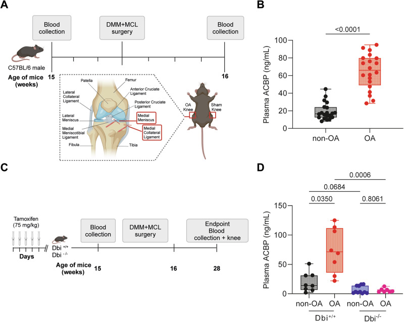
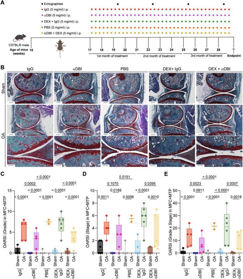
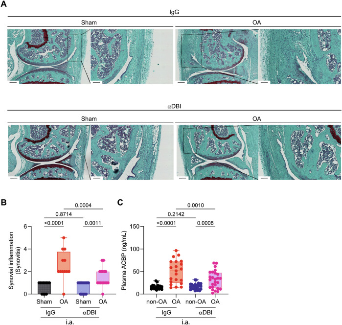
The plasma concentrations of acyl CoA binding protein (ACBP) encoded by the gene diazepam binding inhibitor (DBI) are increased in patients with severe osteoarthritis (OA). Here, we show that knee OA induces a surge in plasma ACBP/DBI in mice subjected to surgical destabilization of one hind limb. Knockout of the Dbi gene or intraperitoneal (i.p.) injection of a monoclonal antibody (mAb) neutralizing ACBP/DBI attenuates OA progression in this model, supporting a pathogenic role for ACBP/DBI in OA. Furthermore, anti-ACBP/DBI mAb was also effective against OA after its intraarticular (i.a.) injection, as monitored by sonography, revealing the capacity of ACBP/DBI to locally reduce knee inflammation over time. In addition, i.a. anti-ACBP/DBI mAb improved functional outcomes, as indicated by the reduced weight imbalance caused by OA. At the anatomopathological level, i.a. anti-ACBP/DBI mAb mitigated histological signs of joint destruction and synovial inflammation. Of note, i.a. anti-ACBP/DBI mAb blunted the OA-induced surge of plasma ACBP/DBI, as well as that of other inflammatory factors including interleukin-1α, interleukin-33, and tumor necrosis factor. These findings are potentially translatable to OA patients because joints from OA patients express both ACBP/DBI and its receptor GABAARγ2. Moreover, a novel mAb against ACBP/DBI recognizing an epitope conserved between human and mouse ACBP/DBI demonstrated similar efficacy in mitigating OA as an anti-mouse ACBP/DBI-only mAb. In conclusion, ACBP/DBI might constitute a promising therapeutic target for the treatment of OA.


Conflict of interest statement

Competing interests: IM is a consultant for Osasuna Therapeutics. GK has been holding research contracts with Daiichi Sankyo, Eleor, Kaleido, Lytix Pharma, PharmaMar, Osasuna Therapeutics, Samsara Therapeutics, Sanofi, Sutro, Tollys, and Vascage. GK is on the Board of Directors of the Bristol Myers Squibb Foundation France. GK is a scientific co-founder of everImmune, Osasuna Therapeutics, Samsara Therapeutics, and Therafast Bio. GK is in the scientific advisory boards of Hevolution, Institut Servier, Longevity Vision Funds, and Rejuveron Life Sciences. GK is the inventor of patents covering therapeutic targeting of aging, cancer, cystic fibrosis, and metabolic disorders. GK’s wife, Laurence Zitvogel, has held research contracts with Glaxo Smyth Kline, Incyte, Lytix, Kaleido, Innovate Pharma, Daiichi Sankyo, Pilege, Merus, Transgene, 9m, Tusk, and Roche, was on the Board of Directors of Transgene, is a co-founder of EverImmune, and holds patents covering the treatment of cancer and the therapeutic manipulation of the microbiota. GK’s brother, Romano Kroemer, was an employee of Sanofi and now consults for Boehringer-Ingelheim. The funders had no role in the design of the study; in the writing of the manuscript, or in the decision to publish the results.

Figures

Fig. 1
Fig. 1. OA-induced increase in plasma ACBP/DBI levels in wild-type mice.
A Schematic representation of the experimental design for osteoarthritis (OA) induction. OA was induced in 15-week-old male mice. Blood samples were collected both before and 1 week after OA induction to assess plasma ACBP/DBI levels. B Plasma ACBP/DBI results are displayed as box-and-whisker plots, with each dot representing an individual mouse (n = 20 mice per condition). For statistical analysis, p values were calculated using a Mann–Whitney test. C Schematic representation of the experimental design for osteoarthritis (OA) induction in ACBP/DBI-deficient mice. D Plasma ACBP/DBI results are displayed as box-and-whisker plots, with each dot representing an individual mouse (n = 8 to 11 mice per condition). For statistical analysis, p values were calculated using a Kruskal–Wallis test. Schematics in (A, C) were generated using BioRender.
Fig. 2
Fig. 2. ACBP/DBI deficiency regulated cartilage degradation in osteoarthritis mice.
A Representative Safranin O-Fast Green and ACBP/DBI-stained knee sections from wild-type and ACBP/DBI-deficient mice. Scale bar, 200 and 50 μm. B Quantification of ACBP/DBI-positive cells relative to total cells in the medial femoral condyle (MFC) and medial tibial plateau (MTP) was presented. Results are displayed as box-and-whisker plots, with each dot representing an individual mouse (n = 4). For statistical analysis, p values were calculated using two-way ANOVA corrected for multiple comparisons. C OARSI semiquantitative scoring for cartilage degradation (grade), D OARSI semiquantitative scoring for cartilage damage (stage), and E a combined index of grade and stage were used to evaluate the extent of cartilage damage. Results are displayed as box-and-whisker plots, with each dot representing an individual mouse (n = 4–8 mice per condition). For statistical analysis, p values were extracted from 2-way linear models, testing surgery significance within different genotypes (formula: Count ~ Genotype/Surgery) and testing genotype significance within surgery status (formula: Count ~ Surgery/Genotype).
Fig. 3
Fig. 3. ACBP/DBI inhibition via monoclonal antibody treatment mitigated osteoarthritis severity.
A Schematic representation of the experimental timeline. In 15-week-old male C57Bl/6 mice, mechanical destabilization of the right knee joint was induced via MCL/DMM surgery, while the left knee underwent sham surgery. After a 2-week post-surgical recovery, OA mice were intraperitoneally injected with either a monoclonal antibody (7G4a) that neutralizes extracellular ACBP/DBI (α-DBI) or an isotype control IgG antibody, administered thrice weekly. This treatment was optionally combined with the synthetic glucocorticoid dexamethasone (DEX). Ultrasound was performed every 3 weeks to monitor inflammation in the joint capsule. Schematic was generated using BioRender. B Representative Safranin O-Fast Green stained knee sections from different treatment groups, illustrating cartilage integrity are shown. Scale bars, 250 and 100 μm. BE Histological evaluation of OA severity. C OARSI semiquantitative scoring for cartilage degradation (grade), D OARSI semiquantitative scoring for cartilage damage (stage), and E a combined index of grade and stage were used to evaluate the extent of cartilage damage. Results are displayed as box-and-whisker plots, with each dot representing an individual mouse (n = 2–6 mice per condition). For statistical analysis, p values were extracted from 2-way linear models, testing treatment significance within different genotypes (formula: Count ~ Genotype/Treatment) and testing genotype significance within different treatments (formula: Count ~ Treatment/Genotype).
Fig. 4
Fig. 4. Effect of α-DBI intraarticular injections on cartilage integrity and weight-bearing asymmetry in OA Mice.
A Schematic representation of the experimental timeline for intraarticular (i.a.) injections of α-DBI and preclinical monitoring in C57Bl/6 OA mice. In 15-week-old male C57Bl/6 mice, mechanical destabilization of the right knee joint was induced via MCL/DMM surgery, while the left knee underwent sham surgery. After a 2-week post-surgical recovery, i.a. injections of α-DBI or isotype control antibody were administered twice weekly for 12 weeks. To manage preclinical follow-up, postural balance measurement began at 16 weeks of age weekly and ultrasound scans were performed every 3 weeks. The mice were euthanized at 28 weeks of age. Schematic was generated using BioRender. Representative knee sections stained for ACBP/DBI (B) and LC3 (D). Scale bar, 50 μm. Quantification of ACBP/DBI-positive cells (C) and LC3-positive cells (E) in the medial femoral condyle (MFC) and medial tibial plateau (MTP). Results are displayed as box-and-whisker plots, with each dot representing an individual mouse (n = 5 mice per condition). For statistical analysis, p values were extracted from 2-way linear models, testing treatment significance within surgery status (formula: Count ~ Surgery/Treatment) and testing surgery significance within different treatments (formula: Count ~ Treatment/Surgery). Weight-bearing asymmetry (F) and the standard deviation of weight-bearing distribution (G) were evaluated over a 3-s period. Ten measurements were taken per mouse, and data are presented as the percentage ratio between weight-bearing on the right and left limbs (F) and the standard deviation of the ratio between the right and left knees (G). Mean ± SEM values (n = 10–13 mice per condition) are shown. Statistical analysis was performed using the area under the curve (AUC), and p values were calculated using ANOVA followed by Tukey’s test for multiple comparisons.
Fig. 5
Fig. 5. ACBP/DBI neutralization improves radiological signs of osteoarthritis, as determined by ultrasound biomicroscope B-mode.
A Echographies of knees after 3,6,9,12 weeks of treatment with IgG and αACBP/DBI mAb clone 7G4a. Tibio-femoral triangle of sham and OA knees was taken using the ultrasound biomicroscope (UBM) in B mode. The femur and tibia are represented in red letters and hypoechogenic zones with a yellow dashed line. B Inflammation kinetic analysis according to the UBM score is shown. Results are displayed as box-and-whisker plots, with each dot representing an individual mouse (n = 9–10 mice per condition). For statistical analysis, P values were extracted from 2-way linear models, testing treatment significance within the week number of treatment (formula: Count ~ Week/Treatment).
Fig. 6
Fig. 6. Intraarticular injection of anti-ACBP/DBI monoclonal antibody reduced cartilage destruction in osteoarthritis.
A Representative Safranin O-Fast Green stained knee sections from mice treated with α-DBI or isotype control antibody. MCL/DMM causes erosion of calcified cartilage extending over 50% of the articular surface in the femur area. Furthermore, the damage starts to extend done the anterior part of the tibial area. In contrast, α-DBI attenuates cartilage loss. Black arrows indicate cartilage lesion. Scale bars 250 and 100 μm. B Semiquantitative scoring system about histological changes. The minimum value 0 corresponds to normal cartilage and the maximum value 12 represents the sum of the destroyed cartilage in MFC and MTP. C Represents the area occupied by the damage in the cartilage, where the value 0 is intact cartilage and the value 8 is the maximum, corresponding to the sum MFC + MTP. D Represents the relationship between the damage and the occupied area. The value 0 is the minimum and 48 is the maximum. Results are displayed as box-and-whisker plots, with each dot representing an individual mouse (n = 9–14 mice per condition). For statistical analysis, p values were extracted from 2-way linear models, testing treatment significance within surgery status (formula: Count ~ Surgery/Treatment) and testing surgery significance within different treatments (formula: Count ~ Treatment/Surgery).
Fig. 7
Fig. 7. ACBP/DBI neutralization reduces synovial inflammation in osteoarthritis mice.
A Representative images of knee synovial membrane from mice treated with α-DBI or isotype control antibody, stained with Safranin O. Scale bars 250 and 100 μm. B Quantification of Krenn score to determine synovitis. Data for each mouse range from 0 (no synovitis) to 9 (maximal inflammation) (n = 12–16 mice per group). For statistical analysis, p values were extracted from 2-way linear models, testing treatment significance within surgery status (formula: Count ~ Surgery/Treatment) and testing surgery significance within different treatments (formula: Count ~ Treatment/Surgery). C Plasma ACBP/DBI levels in Sham and OA mice treated by i.a. injections. Results are displayed as box-and-whisker plots, with each dot representing an individual mouse (n = 9–26 mice per condition). For statistical analysis, p values were calculated using two-way ANOVA corrected for multiple comparisons.
Fig. 8
Fig. 8. ACBP/DBI neutralization reduces pro-inflammatory cytokines in osteoarthritis mice.
A Heatmap representation of the pro-inflammatory cytokine panel in mouse plasma treated with α-DBI or isotype control antibody. The normality of log2-transformed values was tested using the Shapiro–Wilk test. Cytokines with a normal distribution were analyzed by two-way ANOVA with Tukey’s HSD for pairwise comparisons. Non-normally distributed cytokines were analyzed using the Kruskal–Wallis test, followed by Dunn’s post-hoc test with Benjamini-–Hochberg correction for multiple comparisons. Individual representation of cytokine levels for (B) IL1α, (C) IL33, and (D) TNFα. Results are displayed as column plots, with each dot representing an individual mouse (n = 10 mice per condition), shown as mean ± SEM. For statistical analysis, p values were calculated using two-way ANOVA followed by Tukey’s test for multiple comparisons.

References

    1. Safiri S, Kolahi AA, Smith E, Hill C, Bettampadi D, Mansournia MA, et al. Global, regional and national burden of osteoarthritis 1990-2017: a systematic analysis of the Global Burden of Disease Study 2017. Ann Rheum Dis. 2020;79:819–28. - PubMed
    1. Kraus VB, Blanco FJ, Englund M, Karsdal MA, Lohmander LS. Call for standardized definitions of osteoarthritis and risk stratification for clinical trials and clinical use. Osteoarthr Cartil. 2015;23:1233–41. - PMC - PubMed
    1. Loeser RF, Goldring SR, Scanzello CR, Goldring MB. Osteoarthritis: a disease of the joint as an organ. Arthritis Rheum. 2012;64:1697–707. - PMC - PubMed
    1. Sharma L. Osteoarthritis of the knee. N Engl J Med. 2021;384:51–9. - PubMed
    1. Holden MA, Hattle M, Runhaar J, Riley RD, Healey EL, Quicke J, et al. Moderators of the effect of therapeutic exercise for knee and hip osteoarthritis: a systematic review and individual participant data meta-analysis. Lancet Rheumatol. 2023;5:e386–400. - PubMed

MeSH terms

Substances