. 2025 May;44(9):2501-2513.
doi: 10.1038/s44318-025-00408-1. Epub 2025 Mar 13.

An asymmetric nautilus-like HflK/C assembly controls FtsH proteolysis of membrane proteins

Affiliations

An asymmetric nautilus-like HflK/C assembly controls FtsH proteolysis of membrane proteins

Alireza Ghanbarpour et al. EMBO J. 2025 May.

Abstract

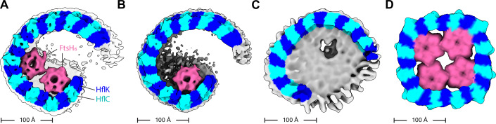
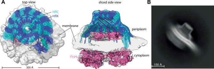
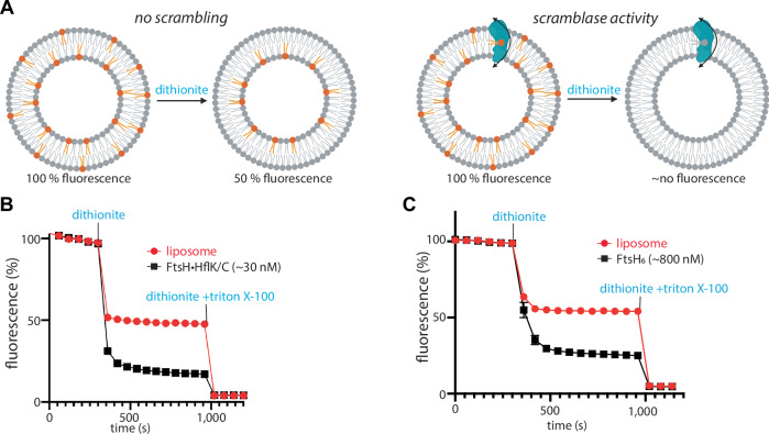
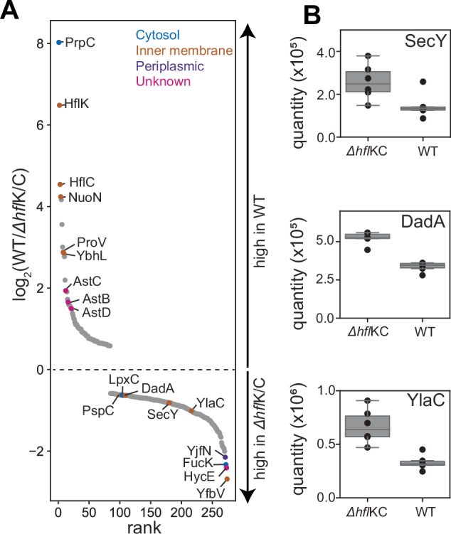
The AAA protease FtsH associates with HflK/C subunits to form a megadalton-size complex that spans the inner membrane and extends into the periplasm of E. coli. How this bacterial complex and homologous assemblies in eukaryotic organelles recruit, extract, and degrade membrane-embedded substrates is unclear. Following the overproduction of protein components, recent cryo-EM structures showed symmetric HflK/C cages surrounding FtsH in a manner proposed to inhibit the degradation of membrane-embedded substrates. Here, we present structures of native protein complexes, in which HflK/C instead forms an asymmetric nautilus-shaped assembly with an entryway for membrane-embedded substrates to reach and be engaged by FtsH. Consistent with this nautilus-like structure, proteomic assays suggest that HflK/C enhances FtsH degradation of certain membrane-embedded substrates. Membrane curvature in our FtsH•HflK/C complexes is opposite that of surrounding membrane regions, a property that correlates with lipid scramblase activity and possibly with FtsH's function in the degradation of membrane-embedded proteins.

Keywords: AAA Protease; Cryo-EM; Macromolecular Complexes; Proteostasis.


Conflict of interest statement

Disclosure and competing interests statement. The authors declare no competing interests.

Figures

Figure 1
Figure 1. Nautilus-like structure of a FtsH•HflK/C super-complex.
(A) Density map (unsharpened) and cartoon models of HflK (blue) and HflC (cyan) viewed from top. Nautilus opening into the HflK/C chamber noted. Map was resolved to a GS-FSC resolution of ~4.5 Å. (B) A sliced top view of the complex highlighting two FtsH hexamers (pink) interacting with SPFH domains of HflK within the nautilus chamber. (C) A side view of the nautilus super-complex, following representation from (A). Approximate location of the membrane depicted with periplasmic and cytoplasmic faces of the inner membrane noted. (D) A hat-like portion of the structure locally refined to ~3.5 Å GS-FSC resolution, color schemes as in (A). This map allowed for clear identification of HflK and HflC. The inset displays is a sharpened map and the model built using this map, highlighting bulky residues in helical regions of HflK and HflC.
Figure 2
Figure 2. HflK/C structures with different numbers of FtsH hexamers.
Density maps were low-pass filtered to 10 Å and colored using the underlying atomic model (HflK—blue; HflC—cyan, FtsH—pink; unassigned membrane or detergent—gray). (A) Two FtsH hexamers per super-complex as refined in Appendix Fig. S10. (B) One FtsH hexamer per super-complex as refined in Appendix Fig. S9. (C) FtsH-free super-complex affinity-purified from cells overexpressing HflC-FLAG and HflK, and refined as depicted in Appendix Figs. S11 and S12. (D) C4-symmetric structure with four FtsH hexamers (Qiao et al, 2022).
Figure 3
Figure 3. Nautilus-like FtsH•HflK/C structure isolated with native lipids via detergent-free solubilization.
(A) Density map highlighting the nautilus FtsH•HflK/C structure determined using affinity purification following detergent-free solubilization using Carboxy-DIBMA (see “Methods”). Map is color-coded (HflK, blue; HflC cyan; FtsH, pink) using docked atomic model, and displayed from the top (left) and as a sliced side view (right). Key structural elements and scale bar noted. (B) 2D-class average of the Carboxy-DIBMA-solubilized sample highlighting membrane reshaping, as evidenced by the belt-like membrane structure that exhibits dual curvature as it traverses through and extends beyond the particle.
Figure 4
Figure 4. FtsH-catalyzed lipid scramblase activity.
(A) Schematic representation of scramblase activity assay, with expected fluorescence level in the absence (left) or presence (right) of enzymatic lipid scrambling activity. Scramblase activity assay of the FtsH•HflK/C complex (B) or isolated FtsH6 (C) using NBD-PE as a substrate. Note that reported protein concentrations do not consider differences in proteoliposome reconstitution efficiency between samples, and that not all liposomes necessarily contain protein. Control reactions assessing the FtsH-dependence of the measured scramblase activity include: (1) a lack of scramblase activity using detergent-solubilized GlpG membrane protein (Ghanbarpour et al, 2021); and (2) NBD-glucose and BSA-back-extraction assays showing that the measured scramblase activity was not a product of partially disrupted, leaky liposomes (Appendix Fig. S15). Values are means (n = 3 technical replicates), with bars marking SEM. Source data are available online for this figure.
Figure 5
Figure 5. Steady-state protein abundance measurements in wild-type and ΔhflK/C cells.
(A) Semi-quantitative proteomic measures of protein abundance in wild-type or ΔhflK/C strains of E. coli. Proteins (n = 274) exhibiting statistically significant changes (false discovery rate q < 0.01, as assessed using an unpaired Student’s t-test corrected for multiple hypothesis testing using a Benjamini–Hochberg procedure implemented in Spectronaut) across six replicates are plotted following the measured rank order abundance change between cell types. A subset of proteins are labeled by gene name, and colored by their annotated localization. Among the highlighted ΔhflK/C-enriched proteins are the putative FtsH substrates LpxC, DadA, SecY, and YlaC. (B) Signal intensity (quantified using label-free precursor ion intensities with Spectronaut) compared between wild-type and ΔhflK/C cells. Inner-quartile range (IQR) depicted by box plot, with dots marking abundance measured in each of six replicates, and with whiskers extending 1.5*IQR. Protein levels changes were statistically significant, with multiple hypothesis testing corrected q values of 7.9 × 10−5 (SecY), 3.6 × 10−11 (DadA), and 8.2 × 10−7 (YlaC).

Update of

References

    1. Amberg-Johnson K, Hari SB, Ganesan SM, Lorenzi HA, Sauer RT, Niles JC, Yeh E (2017) Small molecule inhibition of apicomplexan FtsH1 disrupts plastid biogenesis in human pathogens. eLife 6:e29865 - PMC - PubMed
    1. Arends J, Thomanek N, Kuhlmann K, Marcus K, Narberhaus F (2016) In vivo trapping of FtsH substrates by label-free quantitative proteomics. Proteomics 16:3161–3172 - PubMed
    1. Baker TA, Sauer RT (2012) ClpXP, an ATP-powered unfolding and protein-degradation machine. Biochimica et Biophysica Acta (BBA) Mol Cell Res 1823:15–28 - PMC - PubMed
    1. Bittner LM, Arends J, Narberhaus F (2017) When, how and why? Regulated proteolysis by the essential FtsH protease in Escherichia coli. Biol Chem 398:625–635 - PubMed
    1. Browman DT, Hoegg MB, Robbins SM (2007) The SPFH domain-containing proteins: more than lipid raft markers. Trends Cell Biol 17:394–402 - PubMed

MeSH terms

LinkOut - more resources