

Brain Morphometrics Correlations With Age Among 350 Participants Imaged With Both 3T and 7T MRI: 7T Improves Statistical Power and Reduces Required Sample Size
- PMID: 40083197
- PMCID: PMC11907059
- DOI: 10.1002/hbm.70195
Brain Morphometrics Correlations With Age Among 350 Participants Imaged With Both 3T and 7T MRI: 7T Improves Statistical Power and Reduces Required Sample Size
Abstract
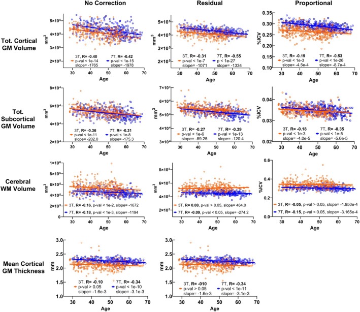
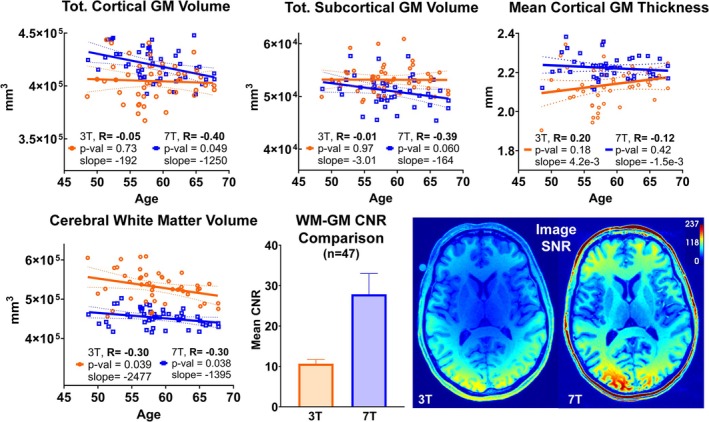
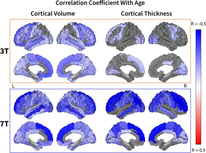
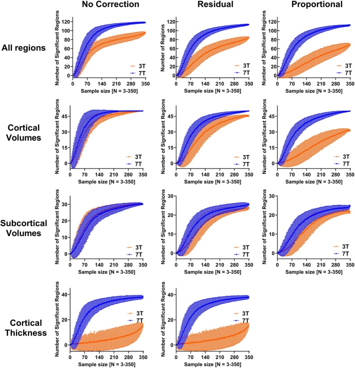
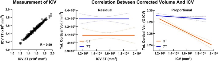
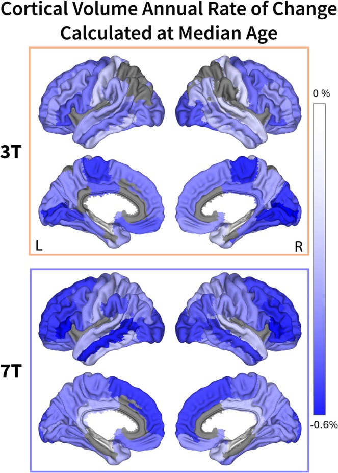
Magnetic resonance imaging (MRI) at 7T has a superior signal-to-noise ratio to 3T but also presents higher signal inhomogeneities and geometric distortions. A key knowledge gap is to robustly investigate the sensitivity and accuracy of 3T and 7T MRI in assessing brain morphometrics. This study aims to (a) aggregate a large number of paired 3T and 7T scans to evaluate their differences in quantitative brain morphological assessment using a widely available brain segmentation tool, FreeSurfer, as well as to (b) examine the impact of normalization methods for subject variability and smaller sample sizes on data analysis. A total of 401 healthy participants aged 29-68 were imaged at both 3T and 7T. Structural T1-weighted magnetization-prepared rapid gradient-echo (MPRAGE) images were processed and segmented using FreeSurfer. To account for head size variability, the brain volumes underwent intracranial volume (ICV) correction using the Residual (regression model) and Proportional (simple division to ICV) methods. The resulting volumes and thicknesses were correlated with age using Pearson's correlation and false discovery rate correction. The correlations were also calculated in increasing sample size from three to the whole sample to estimate the sample size required to detect aging-related brain variation. Three hundred and fifty subjects (208 females) passed the image quality control, with 51 subjects excluded due to excessive motion artifacts on 3T, 7T, or both. 7T MRI showed an overall stronger correlation between morphometrics and age and a larger number of significantly correlated brain volumes and cortical thicknesses. While the ICV is consistent between both field strengths, the Residual normalization method shows markedly higher correlation with age for 3T when compared with the Proportional normalization method. The 7T results are consistent regardless of the normalization method used. In a large cohort of healthy participants with paired 3T and 7T scans, we compared the statistical performance in assessing age-related brain morphological changes. Our study reaffirmed the inverse correlation between brain volumes and cortical thicknesses and age and highlighted varying correlations in different brain regions and normalization methods at 3T and 7T. 7T imaging significantly improves statistical power and thus reduces the required sample size.
Keywords: 3T; 7T; aging; brain morphometrics; magnetic resonance imaging.
© 2025 The Author(s). Human Brain Mapping published by Wiley Periodicals LLC.
Conflict of interest statement
The authors declare no conflicts of interest.
Figures
Update of
-
Brain morphometrics correlations with age among 352 participants imaged with both 3T and 7T MRI: 7T improves statistical power and reduces required sample size.medRxiv [Preprint]. 2024 Nov 1:2024.10.28.24316292. doi: 10.1101/2024.10.28.24316292. medRxiv. 2024. Update in: Hum Brain Mapp. 2025 Mar;46(4):e70195. doi: 10.1002/hbm.70195. PMID: 39574870 Free PMC article. Updated. Preprint.
References
-
- Andrea, N. , Sajewski T. S., De Franco A., et al. 2024. “RF Shimming Strategy for an Open 60‐Channel RF Transmit 7T Head Coil for Routine Use on the Single Transmit Mode.” medRxiv.
-
- Andrea, N. , Sajewski T. S., De Franco A., et al. 2023. “An Open 60‐Channel Tx/ 32‐Channel Rx RF Coil System for Routine Use at 7T ISMRM.”
-
- Benjamini, Y. , and Hochberg Y.. 1995. “Controlling the False Discovery Rate: A Practical and Powerful Approach to Multiple Testing.” Journal of the Royal Statistical Society: Series B: Methodological 57, no. 1: 289–300. http://www.jstor.org.pitt.idm.oclc.org/stable/2346101.
MeSH terms
Grants and funding
- P01 HL040962/HL/NHLBI NIH HHS/United States
- R01 HL105647/HL/NHLBI NIH HHS/United States
- R01MH111265/NH/NIH HHS/United States
- P01HL040962/NH/NIH HHS/United States
- R01 AG053504/AG/NIA NIH HHS/United States
- R01 DK110041/DK/NIDDK NIH HHS/United States
- P01AG025204/NH/NIH HHS/United States
- R01AG053504/NH/NIH HHS/United States
- R01DK110041/NH/NIH HHS/United States
- R56AG074467/NH/NIH HHS/United States
- RF1 AG053504/AG/NIA NIH HHS/United States
- K24 HL123565/HL/NHLBI NIH HHS/United States
- RF1AG053504/NH/NIH HHS/United States
LinkOut - more resources
Full Text Sources
Medical
Miscellaneous
