. 2025 Feb 11;36(1):102482.
doi: 10.1016/j.omtn.2025.102482. eCollection 2025 Mar 11.

Rescue of the disease-associated phenotype in CRISPR-corrected hiPSCs as a therapeutic approach for inherited retinal dystrophies

Affiliations

Rescue of the disease-associated phenotype in CRISPR-corrected hiPSCs as a therapeutic approach for inherited retinal dystrophies

Laura Siles et al. Mol Ther Nucleic Acids. .

Abstract

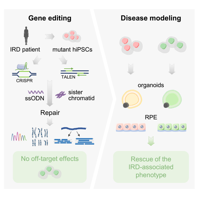
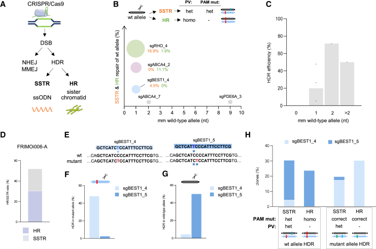
Inherited retinal dystrophies (IRDs), such as retinitis pigmentosa and Stargardt disease, are a group of rare diseases caused by mutations in more than 300 genes that currently have no treatment in most cases. They commonly trigger blindness and other ocular affectations due to retinal cell degeneration. Gene editing has emerged as a promising and powerful strategy for the development of IRD therapies, allowing the permanent correction of pathogenic variants. Using clustered regularly interspaced short palindromic repeats (CRISPR)-Cas9 and transcription activator-like effector nucleases (TALEN) gene-editing tools, we precisely corrected seven hiPS cell lines derived from IRD patients carrying mutations in ABCA4, BEST1, PDE6A, PDE6C, RHO, or USH2A. Homozygous mutations and point insertions/deletions resulted in the highest homology-directed repair efficiencies, with at least half of the clones repaired properly without off-target effects. Strikingly, correction of a heterozygous pathogenic variant was achieved using the wild-type allele of the patient as the template for DNA repair. These results suggest the unexpected potential application of CRISPR as a donor template-free strategy for single-nucleotide modifications. Additionally, the corrected clones exhibited a reversion of the disease-associated phenotype in retinal cellular models. These data strengthen the study and application of gene editing-based approaches for IRD treatment.

Keywords: Best disease; CRISPR; MT: RNA/DNA Editing; TALEN; cell therapy; gene editing; gene therapy; inherited retinal dystrophies; retinitis pigmentosa; sister chromatid.


Conflict of interest statement

The authors declare no conflict of interest.

Figures

None
Graphical abstract
Figure 1
Figure 1
Efficient HDR correction of pathogenic variants in hiPSCs from IRD patients without off-target effects (A) Top: Schematic representation of sgRNAs designed for each pathogenic variant for gene editing in each hiPS cell line. Bottom: Cleavage efficiencies of the sgRNAs (“ND” means not detected). (B) As in (A) but for TALENs. (C) Gene-editing assay overview. The figure was partly generated using Servier Medical Art, provided by Servier, licensed under a Creative Commons Attribution 3.0 Unported license. (D) Gene-editing results for each pathogenic variant. HDR percentages indicate corrected clones without on-target defects from the total clones analyzed. (E) HDR ratios depending on the type of pathogenic variant. Single-nucleotide substitutions refer to mutations in ABCA4_A, BEST1, RHO, and USH2A patients. (F) As in (E) but quantification of on-target aberrations. (G) Bright field captures of hiPS clones after CRISPR-Cas9 (FRIMOi006-A) or TALEN (FRIMOi002-A) gene editing. Scale bar represents 200 μm. (H) Lineage differentiation analysis in FRIMOi002-A parental and corrected clones. Scale bar indicates 50 μm. (I) Table shows the total number of off-targets. Right panel: Scheme of Sanger sequencing screening results (in all hiPS cell lines) and of WGS analysis (in FRIMOi004-A).
Figure 2
Figure 2
Precise HDR correction of pathogenic variants by using the endogenous sister chromatid (A) DNA repair pathways used after CRISPR-Cas9-induced DSB. (B) Percentage of SSTR- and HR-correction in wild-type allele in assays of heterozygous carriers of Figure 1D. (C) Percentage of HDR-mediated correction of the pathogenic variant according to the number of mismatches in homology with the wild-type allele of the sgRNA used. (D) HR and SSTR ratio obtained in the gene-editing assay in FRIMOi006-A. (E) Scheme of sgBEST1_4 and sgBEST1_5 homologies against wild-type and mutant alleles in FRIMOi006-A. Asterisks indicate mismatch in the sequence homology. In red is shown the pathogenic variant and in blue the nucleotide change in the sgRNA. (F) Total HDR efficiency in editing the mutant allele in the assays with sgBEST1_4 or sgBEST1_5 in FRIMOi006-A. (G) As in (F) but in the wild-type allele. (H) HDR-mediated correction data obtained with sgBEST1_4 or sgBEST1_5 according to the pathogenic variant (in green corrected, in red not corrected) and PAM mutation (in blue) in FRIMOi006-A wild-type and mutant alleles.
Figure 3
Figure 3
Pathogenic variant correction by using the wild-type allele as repair template in ssODN-free CRISPR-Cas9 gene-editing assay (A) Venn diagram showing structural variants results found in long-read sequencing in FRIMOi006-A clones. (B) Schematic of the gene-editing assay performed without transfecting the ssODN. (C) Percentage of HR-corrected clones obtained in the assays performed with or without ssODN. (D) Gene-editing ratios based on SSTR or HR repair mechanisms in each hiPS cell line. (E) Representation of percentage of clones with the pathogenic variant corrected in the assays performed without the ssODN depending on the distance between the DSB and the pathogenic variant. (F) Quantification of on-target defects in each assay.
Figure 4
Figure 4
HDR-corrected clones exhibit reversion of the mutant-associated phenotype in retinal cellular models (A) Immunofluorescence staining of ZO-1 or F-actin and RPE65 markers in differentiated RPE FRIMOi006-A clones. Scale bar represents 25 μm. (B) Relative mRNA expression of BEST1 in RPE cells of wild-type and FRIMOi006-A clones. (C) Relative fluorescence quantification of the YFP-labeled Premo Halide Sensor recorded every 30 s for 2 min with the stimulus buffer in RPE cells derived from wild-type, parental, and corrected (SSTR and HR) FRIMOi006-A clones. Data are the average of four independent experiments with different clones of each genotype and represented as mean ± SEM. (D) As in (C) but in the presence of the ionophore A23187. (E) Relative fluorescence units in RPE cultures of parental and corrected FRIMOi006-A clones in the presence of FITC-labeled POS. Data are the average of four independent experiments. (F) Trans-epithelial electrical resistance recordings in wild-type and FRIMOi006-A RPE during cell culture in transwells. Data represent mean ± SEM. p ≤ 0.001 (∗∗∗), p ≤ 0.01 (∗∗), p ≤ 0.05 (∗) levels, or non-significant (ns) for p > 0.05.

References

    1. Tatour Y., Ben-Yosef T. Syndromic inherited retinal diseases: genetic, clinical and diagnostic aspects. Diagnostics. 2020;10 doi: 10.3390/diagnostics10100779. - DOI - PMC - PubMed
    1. Mansfield B.C., Yerxa B.R., Branham K.H. Implementation of a registry and open access genetic testing program for inherited retinal diseases within a non-profit foundation. Am. J. Med. Genet. C Semin. Med. Genet. 2020;184:838–845. doi: 10.1002/ajmg.c.31825. - DOI - PMC - PubMed
    1. Hanany M., Rivolta C., Sharon D. Worldwide carrier frequency and genetic prevalence of autosomal recessive inherited retinal diseases. Proc. Natl. Acad. Sci. USA. 2020;117:2710–2716. doi: 10.1073/pnas.1913179117. - DOI - PMC - PubMed
    1. Schneider N., Sundaresan Y., Gopalakrishnan P., Beryozkin A., Hanany M., Levanon E.Y., Banin E., Ben-Aroya S., Sharon D. Inherited retinal diseases: Linking genes, disease-causing variants, and relevant therapeutic modalities. Prog. Retin. Eye Res. 2022;89 doi: 10.1016/j.preteyeres.2021.101029. - DOI - PubMed
    1. Biswas P., Villanueva A.L., Soto-Hermida A., Duncan J.L., Matsui H., Borooah S., Kurmanov B., Richard G., Khan S.Y., Branham K., et al. Deciphering the genetic architecture and ethnographic distribution of IRD in three ethnic populations by whole genome sequence analysis. PLoS Genet. 2021;17 doi: 10.1371/journal.pgen.1009848. - DOI - PMC - PubMed

LinkOut - more resources