

Management of a patient with arterial thoracic outlet syndrome and Srb anomaly
- PMID: 40083811
- PMCID: PMC11904495
- DOI: 10.1016/j.jvscit.2025.101731
Management of a patient with arterial thoracic outlet syndrome and Srb anomaly
Abstract
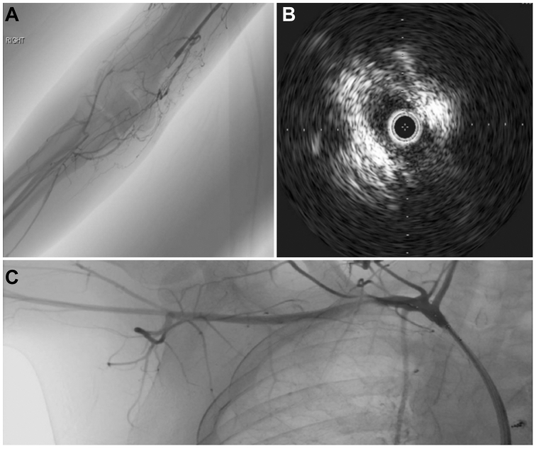
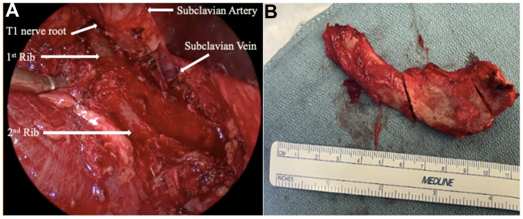
Thoracic outlet syndrome (TOS) is a group of disorders characterized by the compression of neurovascular structures at the thoracic outlet. Arterial TOS, the least common but most severe form, carries significant thromboembolic risks and has a known association with cervical ribs. Synostosis of a complete first and second rib, termed the Srb anomaly, is rare and occurs in approximately 0.2% of the population. Here, we present a unique case of a 17-year-old boy with right upper extremity claudication owing to arterial TOS from an Srb anomaly. This case emphasizes the successful management of an uncommon condition, the importance of accurate diagnosis and timely intervention.
Keywords: Anomalous rib; Rib resection; Thoracic outlet.
© 2025 The Author(s).
Conflict of interest statement
None.
Figures

References
-
- Sanders R.J., Hammond S.L., Rao N.M. Diagnosis of thoracic outlet syndrome. J Vasc Surg. 2007;46:601–604. - PubMed
-
- Vemuri C., McLaughlin L.N., Abuirqeba A.A., Thompson R.W. Clinical presentation and management of arterial thoracic outlet syndrome. J Vasc Surg. 2017;65:1429–1439. - PubMed
-
- Chang K.Z., Likes K., Davis K., Demos J., Freischlag J.A. The significance of cervical ribs in thoracic outlet syndrome. J Vasc Surg. 2013;57:771–775. - PubMed
-
- Oner Z., Oner S., Sahin N.E., Cay M. Evaluation of congenital rib anomalies with multi-detector computed tomography in the Turkish population. Folia Morphol (Warsz) 2024;83:182–191. - PubMed
LinkOut - more resources
Full Text Sources
