HuR inhibition reduces post-ischemic cardiac remodeling by dampening myocyte-dependent inflammatory gene expression and the innate immune response
- PMID: 40085190
- PMCID: PMC11908633
- DOI: 10.1096/fj.202400532RRR
HuR inhibition reduces post-ischemic cardiac remodeling by dampening myocyte-dependent inflammatory gene expression and the innate immune response
Abstract
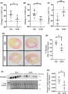
The RNA-binding protein human antigen R (HuR) has been shown to reduce cardiac remodeling following both myocardial infarction and cardiac pressure overload, but the full extent of the HuR-dependent mechanisms within cells of the myocardium has yet to be elucidated. Wild-type mice were subjected to 30 min of cardiac ischemia (via LAD occlusion) and treated with a novel small molecule inhibitor of HuR at the time of reperfusion, followed by direct in vivo assessment of cardiac structure and function. Direct assessment of HuR-dependent mechanisms was done in vitro using neonatal rat ventricular myocytes (NRVMs) and bone marrow-derived macrophages (BMDMs). HuR activity is increased within 2 h after ischemia/reperfusion (I/R) and is necessary for early post-I/R inflammatory gene expression in the myocardium. Despite an early reduction in inflammatory gene expression, HuR inhibition has no effect on initial infarct size at 24 h post-I/R. However, pathological remodeling is reduced with preserved cardiac function at 2 weeks post-I/R upon HuR inhibition. RNA sequencing analysis of gene expression in NRVMs treated with LPS to model damage-associated molecular pattern (DAMP)-mediated activation of toll-like receptors (TLRs) demonstrates a HuR-dependent regulation of pro-inflammatory chemokine and cytokine gene expression in cardiomyocytes. Importantly, we show that conditioned media transfer from NRVMs pre-treated with HuR inhibitor loses the ability to induce inflammatory gene expression and M1-like polarization in bone marrow-derived macrophages (BMDMs) compared to NRVMs treated with LPS alone. Functionally, HuR inhibition reduces macrophage infiltration to the post-ischemic myocardium in vivo. Additionally, we show that LPS-treated NRVMs induce the migration of peripheral blood monocytes in a HuR-dependent endocrine manner. These studies demonstrate that HuR is necessary for early pro-inflammatory gene expression in cardiomyocytes following I/R injury that subsequently mediates monocyte recruitment and macrophage activation in the post-ischemic myocardium.
Keywords: HuR; cardiac remodeling; heart; inflammation; innate immune response; ischemia/reperfusion injury.
© 2025 The Author(s). The FASEB Journal published by Wiley Periodicals LLC on behalf of Federation of American Societies for Experimental Biology.
Conflict of interest statement
The authors state that there is no conflict of interest in connection with this research.
Figures


Update of
-
HuR inhibition reduces post-ischemic cardiac remodeling by dampening acute inflammatory gene expression and the innate immune response.bioRxiv [Preprint]. 2023 Jan 18:2023.01.17.524420. doi: 10.1101/2023.01.17.524420. bioRxiv. 2023. Update in: FASEB J. 2025 Mar 31;39(6):e70433. doi: 10.1096/fj.202400532RRR. PMID: 36711986 Free PMC article. Updated. Preprint.
References
-
- Bacmeister L, Schwarzl M, Warnke S, et al. Inflammation and fibrosis in murine models of heart failure. Basic Res Cardiol. 2019;114:19. - PubMed
MeSH terms
Substances
Grants and funding
- HL166326/HHS | NIH | National Heart, Lung, and Blood Institute (NHLBI)
- 20POST35200267/American Heart Association Postdoctoral Fellowship
- PRE35210795/American Heart Association (AHA)
- HL125204/HHS | NIH | National Heart, Lung, and Blood Institute (NHLBI)
- HL132111/HHS | NIH | National Heart, Lung, and Blood Institute (NHLBI)
- 1029875/American Heart Association (AHA)
- R01 HL166326/HL/NHLBI NIH HHS/United States
- CA252158/HHS | NIH | National Cancer Institute (NCI)
- R01 HL158671/HL/NHLBI NIH HHS/United States
- HL148598/HHS | NIH | National Heart, Lung, and Blood Institute (NHLBI)
- CDA34110117/American Heart Association (AHA)
- CA243445/HHS | NIH | National Cancer Institute (NCI)
- R01 HL132111/HL/NHLBI NIH HHS/United States
- R01 CA243445/CA/NCI NIH HHS/United States
- CA191785/HHS | NIH | National Cancer Institute (NCI)
- 23CDA1052132/American Heart Association Career Development Grant
- F31-HL170636/HHS | NIH | National Heart, Lung, and Blood Institute (NHLBI)
- PRE35230020/American Heart Association (AHA)
- R33 CA252158/CA/NCI NIH HHS/United States
LinkOut - more resources
Full Text Sources
Miscellaneous

