. 2025 Apr 1;214(4):595-605.
doi: 10.1093/jimmun/vkae068.

HLA-E/peptide complexes differentially interact with NKG2A/CD94 and T cell receptors

Affiliations

HLA-E/peptide complexes differentially interact with NKG2A/CD94 and T cell receptors

Linda Voogd et al. J Immunol. .

Abstract

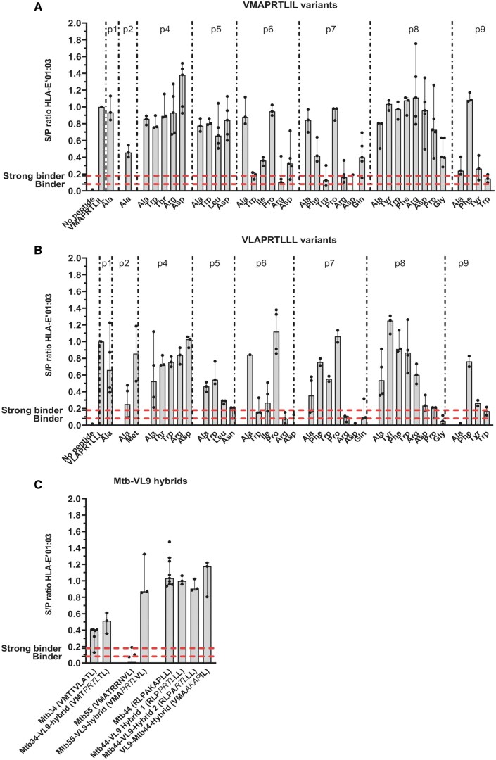
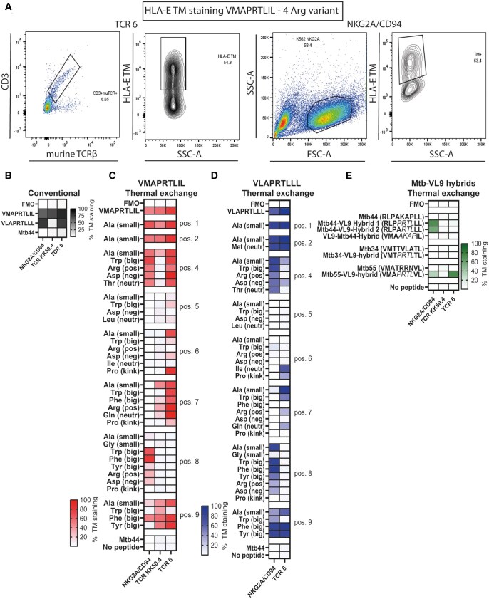
The virtually monomorphic antigen presentation molecule HLA-E can present self- and non-self peptides to the NKG2A/CD94 co-receptor inhibitory complex expressed on natural killer (NK) cells and to T cell receptors (TCRs) expressed on T cells. HLA-E presents self-peptides to NKG2A/CD94 to regulate tissue homeostasis, whereas HLA-E restricted T cells mediate regulatory and cytotoxic responses toward pathogen-infected cells. In this study, we directly compared HLA-E/peptide recognition and signaling between NKG2A/CD94 and 2 HLA-E restricted TCRs that can recognize self-peptides or identical peptide mimics from the viral UL40 protein of cytomegalovirus using position substituted peptide variants. We show that position 7 is critical for interaction with NKG2A/CD94, whereas position 8 is important for interaction with the TCRs. The Arginine at position 5 of these peptides is an essential residue for recognition by both receptors. Thus, NKG2A/CD94 and TCRs have different requirements for recognition of peptides presented in HLA-E.

Keywords: HLA-E; NKG2A/CD94; T cell receptor; peptide/receptor interactions.


Conflict of interest statement

None declared.

Figures

Figure 1.
Figure 1.
Binding of peptide variants and Mtb-VL9 hybrids to HLA-E*01:03. All peptides were evaluated for peptide binding affinity using the previously developed UV-mediated peptide exchange coupled to ELISA assay for HLA-E. (A) Affinity of the VMAPRTLIL variants for HLA-E*01:03. (B) Affinity of the VLAPRTLLL variants for HLA-E*01:03. (C) Affinity of the Mtb-VL9 hybrid peptides for HLA-E*01:03. The sequences of the hybrid peptides are shown on the X-axis and the residues that are changed relative to the wildtype sequence are shown in italics. The residue substitutions at each position are shown on the X-axis in (A) and (B). The Y-axis shows the affinity for HLA-E*01:03, displayed as a signal to positive ratio (S/P) in (A–C). The S/P ratio is a relative measure of the affinity of the peptide variant relative to the positive control peptide (VMAPRTLIL in A and VLAPRTLLL in B). Dotted lines indicate the threshold for defining peptides as binders (>0.08) or good binders (>0.18). These values were defined previously. Peptides were tested at least two times and bars represent the median S/P ratio with 95% confidence interval. Peptide sequences are provided in Table S1.
Figure 2.
Figure 2.
NKG2A/CD94 and TCRs have different requirements for interacting with the HLA-E/peptide complex, localized to positions 4 to 9. K562 cells expressing the NKG2A/CD94 co-receptor (K562-NKG2A/CD94) and Jurkat cells transduced with TCR KK50.4 (recognizing the sequence VMAPRTLIL) and TCR 6 (recognizing VL9 peptides) were stained with HLA-E*01:03 TMs. (A) Representative density plot showing staining with the VMAPRTLIL-Arg 4 variant on Jurkat TCR 6 cells (left) and K562-NKG2A/CD94 cells (right). Jurkat TCR 6 and KK50.4 cells were stained for CD3 and murine TCR-β to gate on the TCR expressing population. (B) Heatmap showing the percentage conventional HLA-E TM staining for the wildtype sequences VMAPRTLIL, VLAPRTLLL and Mtb44 on K562-NKG2A/CD94, Jurkat TCR KK50.4 and TCR 6 cells. (C) Heatmap showing the percentage thermal exchanged HLA-E TM staining on K562-NKG2A/CD94, Jurkat TCR KK50.4 and TCR 6 cells with the VMAPRTLIL variants. (D) Same as C, but then for the VLAPRTLLL variants. (E) Same as C, but then for the Mtb-VL9 hybrid peptides. Residues that were changed in the hybrid peptides relative to the wildtype peptides are shown in italics. Thermal exchanged HLA-E TMs were tested once for binding to NKG2A/CD94, TCR 6 and TCR KK50.4.
Figure 3.
Figure 3.
HLA-E induced signaling of TCR and NKG2A/CD94 depends on the peptide sequence. JE6.1 NKG2A/CD94 NF-kB:eGFP reporter cells and Jurkat TCR KK50.4 triple reporter cells were stimulated with K562 HLA-E*01:03 cells presenting VMAPRTLIL and Mtb-VL9 hybrid peptides. (A) Upper panel: representative plots showing the reporter inhibition signal (eGFP) after stimulating JE6.1 NKG2A/CD94 cells with K562 HLA-E*01:03 cells unloaded with peptide or loaded with Mtb44 (unspecific) or VMAPRTLIL (specific). Reduction of the reporter signal upon peptide stimulation relative to stimulation with unloaded HLA-E*01:03 cells was used to determine the inhibitory signal of the peptide. Lower panel: representative plots showing activation of the triple reporters NFAT (eGFP), AP-1 (mCherry) and NF-kB (eCFP) after stimulating Jurkat TCR KK50.4 cells with K562 HLA-E*01:03 cells loaded with Mtb44 (unspecific) or VMAPRTLIL (specific). (B) Heatmap showing the percentage activation of the TCR triple reporters and percentage inhibition of the NKG2A/CD94 reporter after stimulation with the VMAPRTLIL peptide variants. (C) Same as B, but then for Mtb-VL9 hybrids. Values represent the median percentage reporter activation or inhibition (n = 3 with 2 technical replicates for TCR KK50.4 and n = 2 with 2 technical replicates for NKG2A/CD94).
Figure 4.
Figure 4.
NKG2A/CD94 and TCRs have different recognition and signaling requirements for the HLA-E/peptide complex. (A) Principal component analysis (PCA) on the percentage HLA-E TM staining of the VMAPRTLIL (VM) and VLAPRTLLL (VL) variants on TCR 6 and NKG2A/CD94 expressing cells. Dots are colored based on specificity for either NKG2A/CD94 or TCR 6, shown in the legend. (B) Similar as A, but then between TCR KK50.4 and NKG2A/CD94 expressing cells for the VMAPRTLIL variants. (C) PCA on the reporter signals of TCR KK50.4 and NKG2A/CD94 expressing cells for the VMAPRTLIL variants. (D) Similar as A, but then between TCR KK50.4 and TCR 6 expressing cells for the VMAPRTLIL variants. The top 10% peptides selective for each receptor are named using the single letter amino acid abbreviation, except for (D) as one peptide was recognized by TCR KK50.4. To differentiate between substitutions in the VMAPRTLIL or VLAPRTLLL peptides, the peptide abbreviations in (A). include VM or VL. Substitutions in (B–D) are only in VMAPRTLIL and codes thus refer to peptide position and substitution.

References

    1. Kanevskiy L et al. Dimorphism of HLA-E and its disease association. Int J Mol Sci. 2019;20. - PMC - PubMed
    1. Strong RK et al. HLA-E allelic variants. Correlating differential expression, peptide affinities, crystal structures, and thermal stabilities. J Biol Chem. 2003;278:5082–5090. - PubMed
    1. Grimsley C, Ober C. Population genetic studies of HLA-E: evidence for selection. Hum Immunol. 1997;52:33–40. - PubMed
    1. Joosten SA et al. Mycobacterium tuberculosis peptides presented by HLA-E molecules are targets for human CD8 T-cells with cytotoxic as well as regulatory activity. PLoS Pathog. 2010;6:e1000782. - PMC - PubMed
    1. Prezzemolo T et al. Detailed characterization of human Mycobacterium tuberculosis specific HLA-E restricted CD8(+) T cells. Eur J Immunol. 2018;48:293–305. - PMC - PubMed

MeSH terms

Substances