Multicenter Study
. 2025 Mar 28;27(4):euaf051.
doi: 10.1093/europace/euaf051.

Outcomes of ventricular tachycardia ablation facilitated by pre-procedural cardiac imaging-derived scar characterization: a prospective multi-centre international registry

Affiliations
Multicenter Study

Outcomes of ventricular tachycardia ablation facilitated by pre-procedural cardiac imaging-derived scar characterization: a prospective multi-centre international registry

Diego Penela et al. Europace. .

Abstract

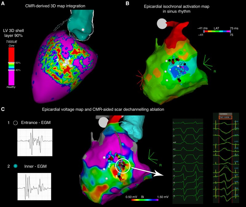
Aims: Pre-procedural imaging can facilitate scar-related ventricular tachycardia (VT) ablation, although only limited data have been reported. This prospective registry aimed to analyse procedural data and outcomes in a multi-centre setting of a pre-defined VT ablation strategy facilitated by the integration of pre-procedural imaging into the navigation system.

Methods and results: Consecutive patients referred for scar-related left-sided VT ablation were prospectively enrolled at five European tertiary hospitals. Pre-procedural cardiac magnetic resonance (CMR)-derived scar maps and/or multi-detector computed tomography (MDCT)-derived wall thinning maps of the left ventricle (LV) were obtained and integrated into the navigation system. An endocardial or endoepicardial approach was chosen based on the scar distribution at pre-procedural imaging. The decision of performing a detailed electro-anatomical map (EAM) of the LV (image-aided) or to using the pre-procedural imaging for guiding the ablation without obtaining an EAM (image-guided) was left to the physician's discretion. One hundred and seventy-one patients (71% with ischaemic cardiomyopathy) were included. Cardiac magnetic resonance was integrated in 159 (93%), MDCT in 113 (66%), and both in 101 (59%) procedures. Procedure-related complications occurred in 9 (5%) patients. At a mean follow-up of 18 ± 19 months, the overall survival and VT recurrence-free survival were 91 and 74.4%, respectively. There were no significant differences in long-term ablation outcomes based on the type of cardiomyopathy (P = 0.88) or the pre-procedural imaging modality employed (P = 0.33). An image-guided approach appears feasible, safe, and faster, with reduced procedure, radiofrequency, and fluoroscopy times, without compromising efficacy.

Conclusion: In a large multi-centre prospective cohort, VT ablation facilitated by pre-procedural imaging is associated with favourable long-term outcomes.

Keywords: Cardiac imaging; Cardiac magnetic resonance; Catheter ablation; Multi-detector computed tomography; Structural heart disease; Substrate ablation; Ventricular tachycardia.


Conflict of interest statement

Conflict of interest: A.Ber. is stockholder of Galgo Medical. D.S.-I. and P.F.-O. are employees of Biosense Webster. P.F. received speaker fees from Boston Scientific and research grants from Abbott and Boston Scientific. All remaining authors have declared no conflicts of interest.

Figures

Graphical Abstract
Graphical Abstract
CMR, cardiac magnetic resonance; LVWT, left ventricle wall thickness; MDCT, multi-detector computed tomography; PSI, pixel signal intensity; VT, ventricular tachycardia.
Figure 1
Figure 1
Cardiac magnetic resonance- and MDCT-derived data post-processing and NSC maps reconstruction of a post-myocardial infarction patient. (A) Pre-procedural MDCT-derived image data post-processing and 3D LV wall thinning map rendering with computed tomography channel detection in a patient with ischaemic cardiomyopathy who experienced an inferior myocardial infarction. (B) Pre-procedural CMR-derived image data post-processing and pixel signal intensity map rendering with BZCs detection in the same patient. LGE-CMR, late gadolinium enhancement-cardiac magnetic resonance; LV, left ventricle; MDCT, multi-detector computed tomography.
Figure 2
Figure 2
Workflow of NSC-derived map integration into the navigation system for ablation procedure. (A) Cardiac magnetic resonance-derived pixel signal intensity map with BZC detection and MDCT-derived LV wall thinning map with computed tomography channel detection superimposition according to standard reference points. (B) The first step of the ablation procedure was the acquisition of an FAM of the aorta in order to integrate MDCT and CMR-derived maps within the spatial reference coordinates of the navigation system. CMR, cardiac magnetic resonance; FAM, fast anatomical map; LVWT, left ventricle wall thinning; MDCT, multi-detector computed tomography; NSC, non-invasive scar characterization; PSI, pixel signal intensity.
Figure 3
Figure 3
Cardiac magnetic resonance-aided epicardial VT ablation in a post-myocarditis patient. (A) Cardiac magnetic resonance-derived PSI map of LV epicardial layer with BZC detection integration into the navigation system. (B) Epicardial isochronal activation map during sinus rhythm and delayed EGM area identification. (C) Voltage EAM rendering and CMR-aided VT exit localization according to pacemapping manoeuvre followed by VT substrate ablation according to scar dechannelling technique, with entrance, and inner channel points RF ablation. CMR, cardiac magnetic resonance; EGM, electrogram; LV, left ventricle.
Figure 4
Figure 4
Cardiac magnetic resonance-guided endocardial VT ablation in a post-myocardial infarction patient with an inferior scar. (A) Cardiac magnetic resonance-guided localization of the arrhythmogenic substrate, classifying EGMs with delayed or fractionated components as entrance, inner, or HSC channel points depending on near-field precocity during sinus rhythm and post-RV extrastimuli. (B) Clinical VT QRS axis-based expected putative channel localization, and RF ablation; all the entrance and inner channel points and HSC-EGMs were targeted for ablation without the realization of an EAM of the LV. CMR, cardiac magnetic resonance; EGM, electrogram; VT, ventricular tachycardia.
Figure 5
Figure 5
Multi-detector computed tomography-aided endocardial VT ablation in a post-myocardial infarction patient with an antero-apical scar. (A) Multi-detector computed tomography-derived LVWT map integration into the navigation system and imaging-aided VT exit localization according to pacemapping. (B) Voltage EAM rendering and VT substrate ablation according to scar dechannelling technique, with entrance, inner channel points, and HSC-EGMs ablation. CC, conducting channel; EGM, electrogram; MDCT, multi-detector computed tomography; VT, ventricular tachycardia.
Figure 6
Figure 6
Procedural outcomes according to the ablation approach.
Figure 7
Figure 7
Ventricular tachycardia recurrence-free survival Kaplan–Meier curve according to the cardiomyopathy diagnosis (A) and according to the ablation approach (B). ICM, ischaemic cardiomyopathy; NICM, non-ischaemic cardiomyopathy; VT, ventricular tachycardia.

References

    1. Reddy VY, Reynolds MR, Neuzil P, Richardson AW, Taborsky M, Jongnarangsin K et al. Prophylactic catheter ablation for the prevention of defibrillator therapy. N Engl J Med 2007;357:2657–65. - PMC - PubMed
    1. Kuck KH, Schaumann A, Eckardt L, Willems S, Ventura R, Delacrétaz E et al. Catheter ablation of stable ventricular tachycardia before defibrillator implantation in patients with coronary heart disease (VTACH): a multicentre randomised controlled trial. Lancet 2010;375:31–40. - PubMed
    1. Arenal A, Avila P, Jimenez-Candil J, Tercedor L, Calvo D, Arribas F et al. Substrate ablation vs antiarrhythmic drug therapy for symptomatic ventricular tachycardia. J Am Coll Cardiol 2022;79:1441–53. - PubMed
    1. Natale A, Zeppenfeld K, Bella PD, Liu X, Sabbag A, Santangeli P et al. Twenty-five years of catheter ablation of ventricular tachycardia: a look back and a look forward. Europace 2023;25:euad225. - PMC - PubMed
    1. Kanagaratnam A, Virk SA, Pham T, Anderson RD, Turnbull S, Campbell T et al. Catheter ablation for ventricular tachycardia in ischaemic versus non-ischaemic cardiomyopathy: a systematic review and meta-analysis. Heart Lung Circ 2022;31:1064–74. - PubMed

Publication types

MeSH terms