. 2025 Jun 24;9(12):2891-2906.
doi: 10.1182/bloodadvances.2024014165.

Endothelial cell activation enhances thromboinflammation in vaccine-induced immune thrombotic thrombocytopenia

Affiliations

Endothelial cell activation enhances thromboinflammation in vaccine-induced immune thrombotic thrombocytopenia

Alexander Dupuy et al. Blood Adv. .

Abstract

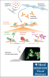
Vaccine-induced immune thrombotic thrombocytopenia (VITT) is a rare but serious complication of the ChAdOx1 nCOV-19 vaccine. In Australia, the diagnosis of VITT required the detection of antibodies against platelet factor 4 (PF4) in plasma using a PF4/polyanion enzyme-linked immunosorbent assay (ELISA). Half of the patients who fulfilled the clinical criteria for VITT tested positive when using this ELISA and another third tested positive when using platelet activation assays, highlighting limitations in the assays used for VITT. Using a microfluidic device coated with endothelial cells, the Endo-chip, we measured the effects of serum and immunoglobulin G (IgG) from patients with clinical VITT on endothelial thromboinflammation. Our cohort comprised 40 patients (21 ELISA-positive and 19 ELISA-negative patients as measured by PF4/polyanion ELISA), 12 vaccinated patients with venous thromboembolism without VITT, and 17 individuals who received the ChAdOx1 vaccine without adverse events (vax controls). Treatment with VITT serum, plasma, or IgG increased endothelial tissue factor (TF) expression and activity. Perfusion of blood from healthy donors labelled with fluorescent antibodies against platelets, neutrophils, and fibrin through Endo-chips treated with VITT serum or IgG induced a twofold to threefold increase in platelet, neutrophil, and fibrin deposition. Thromboinflammation was enhanced with addition of PF4 and reduced with an inhibitory antibody against TF. We conclude that endothelial activation contributes to thromboinflammation in patients with clinical features of VITT. The Endo-chip offers a platform for the study of endothelial responses in immune thrombosis.


Conflict of interest statement

Conflict-of-interest disclosure: The authors declare no competing financial interests.

Figures
None
Graphical abstract
Figure 1.
Figure 1.
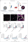
Endo-chip assay. (A) Endothelial cells (HUVECs) are seeded into the microchannel of the chip and allowed to adhere for 4 to 12 hours. Inset on the left shows differential interference contrast image of cells coating the channel. Test sample (eg, patient serum) is injected into the channel and incubated with endothelial cells for 30 minutes and then washed off. Venous blood from a healthy donor is collected into a citrate tube. Fluorescent antibodies are added to the blood (to label platelets, neutrophils, and fibrin). Immediately before perfusion, the whole blood is recalcified. The blood is perfused over the HUVEC layer and fluorescent images are captured by confocal microscopy. Inset on the right shows a representative image of the channel after perfusion of blood showing platelet, fibrin, and neutrophil adhesion to the endothelial layer (thromboinflammation). Image created with BioRender.com. (B) Accumulation of platelets, neutrophils, and fibrin after 15-minute perfusion of blood on HUVECs treated with media, ChAdOx1 nCOV-19, or TNF-α (5 ng/mL, positive control). Representative images of platelet (green), neutrophil (red), and fibrin (magenta) merged images after perfusion of blood. (C) Surface area coverage per field of platelets, number of neutrophils per field, and surface area coverage per field of fibrin at the end of a 15-minute perfusion of blood on HUVECs treated with media, ChAdOx1 nCOV-19, or TNF-α (5 ng/mL). Mean ± standard deviation (SD) of n = 3 independent experiments. One-way analysis of variance (ANOVA) with Tukey post hoc test was used for comparisons. (D) Kinetics of the platelet, neutrophil, and fibrin accumulation on the endothelial surface over 15 minutes of perfusion.
Figure 2.
Figure 2.
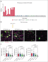
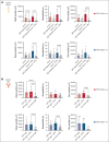
Endothelial thromboinflammation develops in the Endo-chip in response to both VITT ELISA positive and negative sera. (A) Optical density (OD) of VITT samples that tested positive by routine PF4/polyanion ELISA (VITT ELISA positive), and negative by routine PF4/polyanion ELISA (VITT ELISA negative). The OD was also measured for samples from patients with VTE without VITT and in vax controls. The dotted black lines represent the lowest and highest cutoff for positivity determined by the manufacturer. (B) Platelet (green), neutrophil (red), and fibrin (purple) accumulation following a 15-minute blood perfusion in the Endo-chip after treatment with VITT ELISA positive, VITT ELISA negative, VTE without VITT, or vax control sera. Representative images. (C) Platelet fluorescence area per field, neutrophil count per field, and fibrin fluorescence area per field following a 15-minute perfusion of blood in the Endo-chip after treatment with VITT ELISA positive, VITT ELISA negative, VTE without VITT, or vax control sera. VITT ELISA positive n = 15, VITT ELISA negative n = 17, VTE no VITT n = 8, and vax control n = 15. The individual patient numbers are shown in panel A. One-way ANOVA with Dunn post hoc test was used for comparisons.
Figure 3.
Figure 3.
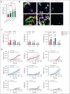
PF4 enhances endothelial thromboinflammation induced by VITT serum in the Endo-chip. (A) Serum concentration of PF4 (ng/mL) in VITT ELISA positive, VITT ELISA negative, VTE without VITT, and vax control samples. One-way ANOVA with Tukey post hoc test was used for comparison. (B) Platelet (green), neutrophil (red), and fibrin (purple) accumulation following a 15-minute blood perfusion in the Endo-chip after treatment with VITT ELISA positive, VITT ELISA negative, or vax control serum without (upper panel) or with (lower panel) the addition of PF4. Representative images. (C) Platelet fluorescence area per field, neutrophil count per field, and fibrin fluorescence area per field following a 15-minute blood perfusion in the Endo-chip after treatment with VITT ELISA positive serum without or with addition of PF4, VITT ELISA negative serum without or with addition of PF4, and vax control serum without or with addition of PF4. A paired t test was used for comparison. (D) Kinetics of the platelet fluorescence area, neutrophil count, and fibrin fluorescence area over 15 minutes of blood perfusion in the Endo-chip after treatment with VITT ELISA positive serum without or with addition of PF4, VITT ELISA negative serum without or with addition of PF4, or vax control serum without or with addition of PF4. VITT ELISA positive n = 7 (patients 1, 2, 4, 5, 6, 8, 11), VITT ELISA negative n = 11 (patients 1, 2, 4, 5, 6, 8, 9, 10, 11, 12, 14), vax controls n = 11 (patients 1, 2, 5, 6, 7, 8, 10, 12, 13, 14, 15). PF4 was added at 25 μg/mL in all experiments.
Figure 4.
Figure 4.
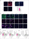
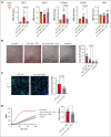
VITT serum induces endothelial cell activation, which is enhanced with the addition of PF4. (A) Immunostaining of the HUVEC layer for spike protein (red), nuclei, TF, and P-selectin (lower panel) after incubation with media alone or with media containing ChAdOx1 nCOV-19 adenoviral vaccine. (B) Immunostaining for human IgG (green) in HUVECs after treatment with VITT sera vs vax control sera. Representative images. The fluorescence intensity of IgG per field is shown for VITT and vax controls. Mean ± SD from 3 to 5 fields of view; n = 4 independent experiments, unpaired t test. (C) Representative images of HUVECs stained for TF (green), P-selectin (red), and VCAM-1 (magenta) after exposure to TNF-α (5 ng/mL), VITT serum, VITT serum + PF4 (25 μg/mL), vax control serum, vax control serum + PF4 (25 μg/mL), or media. Nuclear staining using Hoechst is shown in blue. (D) Fluorescence intensity of TF, P-selectin, and VCAM-1 of HUVECS treated with VITT, without or with 25 μg/mL PF4 added, or treated with vax control, without or with 25 μg/mL PF4 added, expressed as fold change in comparison with the fluorescent intensity of media alone. Mean ± SD, from 3 to 5 fields of view; VITT ELISA positive n = 2 (patients 2, 4), VITT ELISA negative n = 2 (patients 3, 9), vax control n = 4 (patients 2, 6, 10, 12). One-way ANOVA with Dunn post hoc test was used for comparison. (E) Fold change in the mRNA expression of F3 (TF) in HUVECs treated with plasma, with or without PF4 (25 μg/mL), in comparison with that of glyceraldehyde-3-phosphate dehydrogenase. Mean ± SD, n = 10 for media, VITT ELISA positive n = 21 (Table 1), VITT ELISA negative n = 17 (Table 1), vax control n = 8 plasma samples. One-way ANOVA with Dunn post hoc test was used for comparisons.
Figure 5.
Figure 5.
The endothelial thromboinflammatory response in VITT is antibody dependent. (A) Platelet fluorescence area per field, neutrophil count per field, and fibrin fluorescence area per field following a 15-minute blood perfusion in the Endo-chip after treatment with VITT ELISA positive serum or IgG depleted serum (upper panel) or with VITT ELISA negative serum or IgG depleted serum (lower panel) in the presence or absence of 25 μg/mL PF4. VITT ELISA positive n = 5 (patients 2, 3, 4, 6, 12), VITT ELISA negative n = 5 (patients 1, 3, 4, 5, 6). One-way ANOVA with Dunn post hoc test was used for comparisons. (B) Platelet fluorescence area per field, neutrophil count per field, and fibrin fluorescence area per field following a 15-minute blood perfusion in the Endo-chip after treatment with IgG isolated from the same VITT ELISA positive serum or VITT ELISA negative serum as in (A), in the presence or absence of 25 μg/mL PF4, in comparison with IgG isolated from vax control serum. VITT ELISA positive n = 5 (patients 2, 3, 4, 6, 12), VITT ELISA negative n = 5 (patients 1, 3, 4, 5, 6), vax controls n = 5. One-way ANOVA with Dunn post hoc test was used for comparison.
Figure 6.
Figure 6.
Treatment with VITT IgG and PF4 induces endothelial activation. (A) Relative mRNA expression of endothelial cells to glyceraldehyde-3-phosphate dehydrogenase in endothelial cells incubated with media (untreated), treated with VITT IgG and 25 μg/mL PF4, or with vax control IgG and 25 μg/mL PF4. Expression was determined for E-selectin (SELE), P-selectin (SELP), VCAM-1 (VCAM1), ICAM-1 (ICAM1), and VWF. There were n = 3 repeats of n = 3 VITT ELISA positive IgG (patients 3, 4, 11) and n = 3 vax control IgG (patients 1, 2, 5). One-way ANOVA with Dunn post hoc analysis was used for comparisons. (B) Left: release of VWF after stimulation of endothelial cells with thrombin, VITT IgG and 25 μg/mL PF4, vax control IgG and 25 μg/mL PF4, or untreated. Representative images. Right: quantification of the average number of VWF strings per field after stimulation with thrombin, VITT IgG + PF4, vax control IgG + PF4, and untreated. A total of 2 to 3 fields of view were used for n = 3 VITT ELISA positive IgG (patients 3, 4, 11) and n = 3 vax control IgG (patients 1, 2, 5). One-way ANOVA with Tukey post hoc test was used for comparisons. (C) Left: representative immunofluorescent images of TF staining (green) of HUVECs after incubation with IgG isolated from VITT or vax control serum. Nuclei are stained with Hoechst (blue). Right: TF fluorescence intensity units after incubation of HUVECs with VITT IgG or vax control IgG; average of n = 3 fields of view from 4 VITT ELISA positive (patients 2, 4, 6, 13) and 3 vax controls (patients 1, 2, 5). Unpaired t tests were used for comparisons. (D) Left: kinetic curve of Xa generation after treatment of endothelial cells with media (untreated), VITT IgG + 25 μg/mL PF4, vax control IgG + 25 μg/mL PF4, and 10 ng/mL TNF-α (left panel). Right: factor Xa generation after treatment of endothelial cells with VITT IgG + PF4 vs vax control IgG + PF4. There were 3 repeats for 3 VITT ELISA positive IgG samples (patients 4, 6, 13) and 3 vax control IgG samples (patients 1, 2, 5). Unpaired t tests were used for comparison.
Figure 7.
Figure 7.
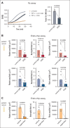
Inhibition of TF and thrombin decreases the endothelial thromboinflammatory response to VITT serum. (A) Left: kinetics of factor Xa generation in endothelial cells after incubation with media (untreated) or TNF-α in the presence of isotype control antibody or inhibitory anti-TF antibody 5G9. Right: factor Xa generation (nanomoles per minute [nM/min]) of TNF-α–treated HUVECs in the presence of isotype control or 5G9 antibody. A paired t test was used for comparisons. (B) Platelet fluorescence area per field, neutrophil count per field, and fibrin fluorescence area per field after a 15-minute blood perfusion in the Endo-chip after treatment with VITT ELISA positive or VITT ELISA negative sera in the presence of isotype control or 5G9 antibody. VITT ELISA positive n = 6 (patients 2, 4, 5, 6, 9, 13), VITT ELISA negative n = 6 (patients 2, 4, 9, 10, 13, 14). A paired t test was used for comparison. (C) Platelet fluorescence area per field, neutrophil count per field, and fibrin fluorescence area per field following 15 minutes perfusion with blood containing vehicle control or the thrombin inhibitor argatroban in the Endo-chip after treatment with VITT serum with 25 μg/mL PF4 added. VITT ELISA positive sera n = 3 (patients 4, 6, 13). A paired t test was used for comparison.

References

    1. Greinacher A, Thiele T, Warkentin TE, Weisser K, Kyrle PA, Eichinger S. Thrombotic thrombocytopenia after ChAdOx1 nCov-19 vaccination. N Engl J Med. 2021;384(22):2092–2101. - PMC - PubMed
    1. Schultz NH, Sørvoll IH, Michelsen AE, et al. Thrombosis and thrombocytopenia after ChAdOx1 nCoV-19 vaccination. N Engl J Med. 2021;384(22):2124–2130. - PMC - PubMed
    1. Scully M, Singh D, Lown R, et al. Pathologic antibodies to platelet factor 4 after ChAdOx1 nCoV-19 vaccination. N Engl J Med. 2021;384(23):2202–2211. - PMC - PubMed
    1. Uzun G, Zlamal J, Althaus K, et al. Cerebral venous sinus thrombosis and thrombocytopenia due to heparin-independent anti-PF4 antibodies after adenovirus infection. Haematologica. 2024;109(6):2010–2015. - PMC - PubMed
    1. Warkentin TE, Baskin-Miller J, Raybould AL, et al. Adenovirus-associated thrombocytopenia, thrombosis, and VITT-like antibodies. N Engl J Med. 2023;389(6):574–577. - PubMed