. 2025 Mar 14;39(7-8):510-523.
doi: 10.1101/gad.352498.124. Online ahead of print.

MdfA is a novel ClpC adaptor protein that functions in the developing Bacillus subtilis spore

Affiliations

MdfA is a novel ClpC adaptor protein that functions in the developing Bacillus subtilis spore

Shawn C Massoni et al. Genes Dev. .

Abstract

Bacterial protein degradation machinery consists of chaperone-protease complexes that play vital roles in bacterial growth and development and have sparked interest as novel antimicrobial targets. ClpC-ClpP (ClpCP) is one such chaperone-protease complex, recruited by adaptors to specific functions in the model bacterium Bacillus subtilis and other Gram-positive bacteria, including the pathogens Staphylococcus aureus and Mycobacterium tuberculosis Here we have identified a new ClpCP adaptor protein, MdfA (metabolic differentiation factor A; formerly YjbA), in a genetic screen for factors that help drive B. subtilis toward metabolic dormancy during spore formation. A knockout of mdfA stimulates gene expression in the developing spore, while aberrant expression of mdfA during vegetative growth is toxic. MdfA binds directly to ClpC to induce its oligomerization and ATPase activity, and this interaction is required for the in vivo effects of mdfA Finally, a cocrystal structure reveals that MdfA binds to the ClpC N-terminal domain at a location analogous to that on the M. tuberculosis ClpC1 protein where bactericidal cyclic peptides bind. Altogether, our data and that of an accompanying study by Riley and colleagues support a model in which MdfA induces ClpCP-mediated degradation of metabolic enzymes in the developing spore, helping drive it toward metabolic dormancy.

Keywords: AAA+ proteases; Bacillus subtilis; ClpC; ClpCP; MdfA; X-ray crystallography; YjbA; adaptor; protein degradation; sporulation.


Figures

Figure 1.
Figure 1.
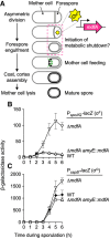
Forespore gene expression is stimulated in cells deleted for mdfA. (A) Cartoon representation of B. subtilis sporulation, with major morphological events indicated. Also shown is the SpoIIQ•SpoIIIAA-AH channel apparatus through which the mother cell provides small molecule metabolites to support the forespore during late development (Camp and Losick 2009; Riley et al. 2021). The early-acting forespore σ factor σF is shown, as is the mdfA (formerly yjbA) gene, which has been assigned to the σF regulon (Steil et al. 2005; Wang et al. 2006; Arrieta-Ortiz et al. 2015) and is the focus of this study. (B,C) The forespore-specific σ factors σF and σG are significantly more active in ΔmdfA cells. Production of β-galactosidase from the σF-dependent PspoIIQ-lacZ reporter (B) or σG-dependent PsspB-lacZ reporter (C) was monitored during sporulation of otherwise wild-type cells (WT; closed diamonds), cells deleted for mdfAmdfA; open inverted triangles), or cells deleted for mdfA and harboring mdfA integrated at the amyE locus (ΔmdfA amyE::mdfA; open triangles; PspoIIQ-lacZ strains AHB1841, CFB530, and CFB541, respectively; PsspB-lacZ strains AHB324, SMB179, and SMB334, respectively). Error bars indicate ±standard deviations based on three or more independent experiments.
Figure 2.
Figure 2.
The clpCQ11P allele suppresses vegetative mdfA toxicity and, like ΔmdfA, stimulates σF activity during sporulation. (A) Expression of mdfA during vegetative growth causes cell death at high inducer concentrations, and this toxicity is suppressed by mutations in clpC and clpP. Wild-type cells and cells harboring an engineered IPTG-inducible mdfA gene construct (WT and PIPTG-mdfA; strains PY79 and CFB189, respectively), as well as PIPTG-mdfA cells harboring clpCQ11P, clpCQ11A, ΔclpC, or ΔclpP (strains SFB32, SFB38, CFB241, and CFB274, respectively), were grown to mid-logarithmic phase (OD600 ∼ 0.5) in LB media and adjusted to OD600 = 1, and 10-fold serial dilutions (10−1–10−4, left to right) were spotted onto LB plates with or without 1 mM IPTG. Plates were imaged after 24 h of growth at 37°C. (B) Expression of mdfA during vegetative growth causes cell filamentation at lower inducer concentrations, and this filamentation is suppressed by clpCQ11P and ΔclpC. WT, PIPTG-mdfA, PIPTG-mdfA clpCQ11P, and PIPTG-mdfA ΔclpC cells (strains PY79, CFB189, SMB361, and CFB241, respectively) were grown for ∼24 h at 37°C on LB agar plates with 50 µM IPTG. Colonies were picked, diluted, and visualized by phase microscopy. Arrowheads indicate filamentous cells that appear to have lysed. Scale bar, 10 µm. (C) Domain structure of the ClpC protein. Shown are the adaptor/substrate-binding N domain (blue), AAA+ ATPase D1 domain (yellow), M domain (magenta), and AAA+ ATPase D2 domain (cyan). The ClpP binding site within the D2 domain is also indicated (green). The location of the Q11 residue in the N domain is labeled. (D) Cells harboring the clpCQ11P allele, unlike those harboring ΔclpC, do not have a severe sporulation defect. WT, ΔclpC, and clpCQ11P cells (strains PY79, CFB270, and CFB282, respectively) were induced to sporulate for 24 h, and the number of heat-resistant spores per milliliter was determined. Individual data points are shown (n = 6), and bars indicate the mean number of heat-resistant spores per milliliter for each strain. (E) The clpCQ11P allele, like ΔmdfA but unlike ΔclpC, significantly stimulates σF activity during sporulation but does not further stimulate σF activity in the absence of mdfA. β-Galactosidase production from the σF-dependent PspoIIQ-lacZ reporter was monitored during sporulation of otherwise wild-type cells (WT; closed diamonds), cells deleted for mdfAmdfA; open inverted triangles), cells deleted for clpCclpC; open squares), cells harboring clpCQ11P (clpCQ11P; solid triangles), or cells deleted for mdfA and harboring clpCQ11PmdfA clpCQ11P; solid circles; strains AHB881, SMB189, CFB302, CFB307, and CFB335, respectively). Error bars indicate ±standard deviations based on four or more replicates.
Figure 3.
Figure 3.
MdfA interacts with the ClpC N domain in a Q11-dependent manner. (A) Isothermal titration calorimetry (ITC) experiments indicate that MdfA and ClpCN (left), but not MdfA and ClpCN,Q11P (right), form a complex in vitro. Twenty injections of 2 μL of MdfA at a concentration of 700 μM were added to ClpC constructs at 70 μM in the reaction cell. The heat of MdfA dilution into buffer was subtracted from the data. Raw data are shown in the top panels, and binding isotherm is shown in the bottom panels. Fitted data for MdfA–ClpCN interaction are as follows: ΔH = −19.5 kJ/mol ± 0.6 kJ/mol, KD = 3.79 µM ± 0.50 µM, and N = 0.82 sites ± 0.01 sites. (B) MdfA and ClpC interact in a transcription-based bacterial two-hybrid assay in a manner that depends on the ClpC N domain and Q11. In this assay, interaction of “bait” and “prey” proteins, fused to the bacteriophage λ cI protein (λcI) and the N-terminal domain of the α subunit of RNA polymerase (α). respectively, drives expression of a lacZ reporter gene under the control of a test promoter in Escherichia coli, leading to production of β-galactosidase (Dove and Hochschild 2004). ClpC or variants thereof was expressed from plasmids as fusions to λcI (“bait”), whereas MdfA or the well-characterized ClpC adaptor protein MecA was expressed as fusions to α (“prey”). The full list of bacterial two-hybrid bait and prey plasmids is in Supplemental Table S2. Variants of ClpC (ClpCND1M, ClpCN, ClpCD1M, and ClpCD2) harboring one or more of its domains and/or the Q11P substitution are depicted in a manner derived from Figure 2C. The Q11P substitution is indicated by a pale-green asterisk, and data collected from pairs including a ClpC Q11P variant are shaded pale green. The dotted line represents background β-galactosidase activity in a negative control strain expressing λCI and the α subunit of RNA polymerase alone (depicted as “—” for both prey and bait). Data are plotted as mean values, with error bars indicating ±standard deviation (n ≥ 5 replicate values). (***) P < 0.0001, unpaired t-test with Welch's correction.
Figure 4.
Figure 4.
MdfA induces oligomerization and ATPase activity of full-length ClpC in vitro but does not direct degradation of a test substrate. (A,B) Like MecA, MdfA induces the formation of stable higher oligomeric complexes with a ClpC double-Walker-B variant (ClpCDWB) in the presence of ATP, as detected by size exclusion chromatography. The elution profiles of all proteins alone (A) and in combinations (B) (as indicated; 10 µM each) were detected by absorbance at 280 nm. The black arrows indicate the positions and molecular weights of native marker proteins: thyroglobulin (669 kDa), ferritin (440 kDa), aldolase (158 kDa), ovalbumin (44 kDa), and carbonic anhydrase (29 kDa). Elution fractions were concentrated and analyzed by SDS-PAGE followed by Coomassie staining, the results of which are shown below each relevant fraction. (C) Like MecA, MdfA induces the ATPase activity of ClpC. The ATPase activity of 1 µM ClpC, 1 µM ClpCQ11P, or 1 µM ClpCQ11A was determined in the presence of 1 µM MecA or 1–15 µM MdfA (as indicated). All ATPase activities were normalized to that of ClpC induced by MecA. Error bars indicate ±standard deviations based on at least three replicates. (D,E) Unlike MecA, MdfA does not induce ClpCP-mediated degradation of itself or α/β-casein. Samples from in vitro degradation reactions harboring the indicated proteins (1 µM each) as well as an ATP regeneration system (pyruvate kinase [PK]) were taken at 0, 30, and 90 min and analyzed by SDS-PAGE followed by Coomassie staining.
Figure 5.
Figure 5.
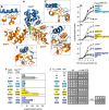
A cocrystal structure of MdfA and ClpCN. (A) Complete X-ray crystal structure of MdfA (orange) in complex with ClpCN (blue), with Q11 highlighted (lime green space fill), solved to 2.0 Å resolution. (BE) Zoomed views of the interface between MdfA (orange) and ClpCN (blue). Three pairs of charged residues predicted to form electrostatic interactions between the two proteins are highlighted: ClpC K12 and MdfA E161, ClpC K85 and MdfA E116, and ClpC H79 and MdfA E53. (F) Mutational analysis of the MdfA-E161/ClpC-K12, MdfA-E116/ClpC-K85, and MdfA-E53/ClpC-H79 electrostatic interactions in MdfA–ClpCND1M binding in the bacterial two-hybrid assay. See the legend for Figure 3 for more details. The full list of bacterial two-hybrid bait and prey plasmids is in Supplemental Table S2. The dotted line represents background β-galactosidase activity in a negative control strain (depicted as “—” for both prey and bait). Data are plotted as mean values, with error bars indicating ±standard deviation (n ≥ 4 replicate values). (GI) Mutational analysis of the MdfA-E161/ClpC-K12 (G), MdfA-E116/ClpC-K85 (H), and MdfA-E53/ClpC-H79 (I) electrostatic interactions during sporulation. β-Galactosidase production from the σF-dependent PspoIIQ-lacZ reporter was monitored during sporulation of otherwise wild-type cells (WT; closed diamonds), cells deleted for mdfAmdfA; open inverted triangles), or cells harboring the indicated mutations in mdfA and/or clpC (closed squares/circles/triangles indicate mdfA single mutants, open squares/circles/triangles indicate clpC single mutants, and shaded squares/circles/triangles indicate mdfA clpC double mutants). Strains used were as follows: WT (AHB881), ΔmdfA (SMB189), mdfAE161K (SYB10), clpCK12E (RFB32), mdfAE161K clpCK12E (AHB6234), mdfAE116K (AHB6220), clpCK85E (AHB6211), mdfAE116K clpCK85E (AHB6229), mdfAE53H (AVB6), clpCH79E (AHB6207), and mdfAE53H clpCH79E (AHB6231). Error bars indicate ±standard deviations based on three or more replicates. The same WT and ΔmdfA data are shown in all three panels for clarity. (J) Mutational analysis of the MdfA-E161/ClpC-K12, MdfA-E116/ClpC-K85, and MdfA-E53/ClpC-H79 electrostatic interactions in MdfA toxicity during vegetative growth. Cells harboring an engineered IPTG-inducible mdfA gene construct (PIPTG-mdfA) with either wild-type (WT) or mutant mdfA alleles (E161K, E116K, or E53H) and harboring either wild-type (WT), deletion (Δ), or other mutant alleles (K12E, K85E, or H79E) of clpC at its native locus were grown for 6 h in LB media at 37°C; subjected to 10-fold serial dilutions (10−2–10−5, left to right); and spotted using a sterile 48 pin custom-made replicator onto LB plates with or without 1 mM IPTG. A wild-type strain without PIPTG-mdfA (—) was also included as a control. Note that the native mdfA gene was unaltered in these strains. Plates were grown at 25°C and imaged after 48 h and, in the case of the bottom far-right panel, 72 h. Note that the PIPTG-mdfAE53H clpCH79E strain shows a small colony phenotype in the presence of inducer and evidence of cell lysis after another 24 h of growth. Strains used were as follows (listed as in the panel from top to bottom): PY79, CFB189, CFB241, CSB8, RFB21, AHB6213, XWB12, XWB50, XWB58, SFB8, SOB18, and AHB6215.
Figure 6.
Figure 6.
MdfA binds ClpCN in a location distinct from that bound by the B. subtilis ClpC adaptor MecA and phosphoarginine (pArg) peptides but similar to the surface of M. tuberculosis ClpC1N bound by the antibiotics cyclomarin A, ecumicin, and rufomycin. (AC) Cocrystal structures of the B. subtilis ClpC N domain bound to MdfA (A) (PDB: 8B3S; this study), MecA (B) (PDB: 3PXG; Wang et al. 2011), and phosphoarginine (pArg; C) (PDB: 5HBN; Trentini et al. 2016). (DF) Cocrystal structures of the M. tuberculosis ClpC N domain bound to cyclomarin A (D) (PDB: 3WDC; Vasudevan et al. 2013), ecumicin (E) (PDB: 6PBS; Wolf et al. 2020), and rufomycin (F) (PDB: 6CN8; Wolf et al. 2019).
Figure 7.
Figure 7.
Modeling MdfA onto the ClpC hexamer indicates a potential steric clash with the ClpC M domain. (A) Cocrystal structure (PDB: 3J3T) of B. subtilis MecA (green) in a hexameric complex with ClpC (gray) (Liu et al. 2013). (B,C) Alternate views of the MdfA/ClpCN crystal complex (PDB: 8B3S; this study) aligned with ClpCN in the full-length ClpC hexamer shown in A, indicating that MdfA (orange) and MecA (green) occupy different binding positions on ClpCN. (D) Zoomed view showing the steric clash between MdfA (orange) and the ClpC M domain (magenta). The overlay of ClpCN from the hexamer (gray) and the MdfA/ClpCN cocrystal structure (blue) is shown in D but removed from B and C for clarity.

References

    1. Arrieta-Ortiz ML, Hafemeister C, Bate AR, Chu T, Greenfield A, Shuster B, Barry SN, Gallitto M, Liu B, Kacmarczyk T, et al. 2015. An experimentally supported model of the Bacillus subtilis global transcriptional regulatory network. Mol Syst Biol 11: 839. 10.15252/msb.20156236 - DOI - PMC - PubMed
    1. Camp AH, Losick R. 2009. A feeding tube model for activation of a cell-specific transcription factor during sporulation in Bacillus subtilis. Genes Dev 23: 1014–1024. 10.1101/gad.1781709 - DOI - PMC - PubMed
    1. Carroni M, Franke KB, Maurer M, Jäger J, Hantke I, Gloge F, Linder D, Gremer S, Turgay K, Bukau B, et al. 2017. Regulatory coiled-coil domains promote head-to-head assemblies of AAA+ chaperones essential for tunable activity control. eLife 6: e30120. 10.7554/eLife.30120 - DOI - PMC - PubMed
    1. Choules MP, Wolf NM, Lee H, Anderson JR, Grzelak EM, Wang Y, Ma R, Gao W, McAlpine JB, Jin Y-Y, et al. 2019. Rufomycin targets ClpC1 proteolysis in Mycobacterium tuberculosis and M. abscessus. Antimicrob Agents Chemother 63: e02204-18. 10.1128/AAC.02204-18 - DOI - PMC - PubMed
    1. Collins GA, Goldberg AL. 2017. The logic of the 26S proteasome. Cell 169: 792–806. 10.1016/j.cell.2017.04.023 - DOI - PMC - PubMed

LinkOut - more resources