. 2025 Apr;120(2):373-392.
doi: 10.1007/s00395-025-01101-4. Epub 2025 Mar 15.

Necrosis-like cell death modes in heart failure: the influence of aetiology and the effects of RIP3 inhibition

Affiliations

Necrosis-like cell death modes in heart failure: the influence of aetiology and the effects of RIP3 inhibition

Izabela Jarabicová et al. Basic Res Cardiol. 2025 Apr.

Abstract

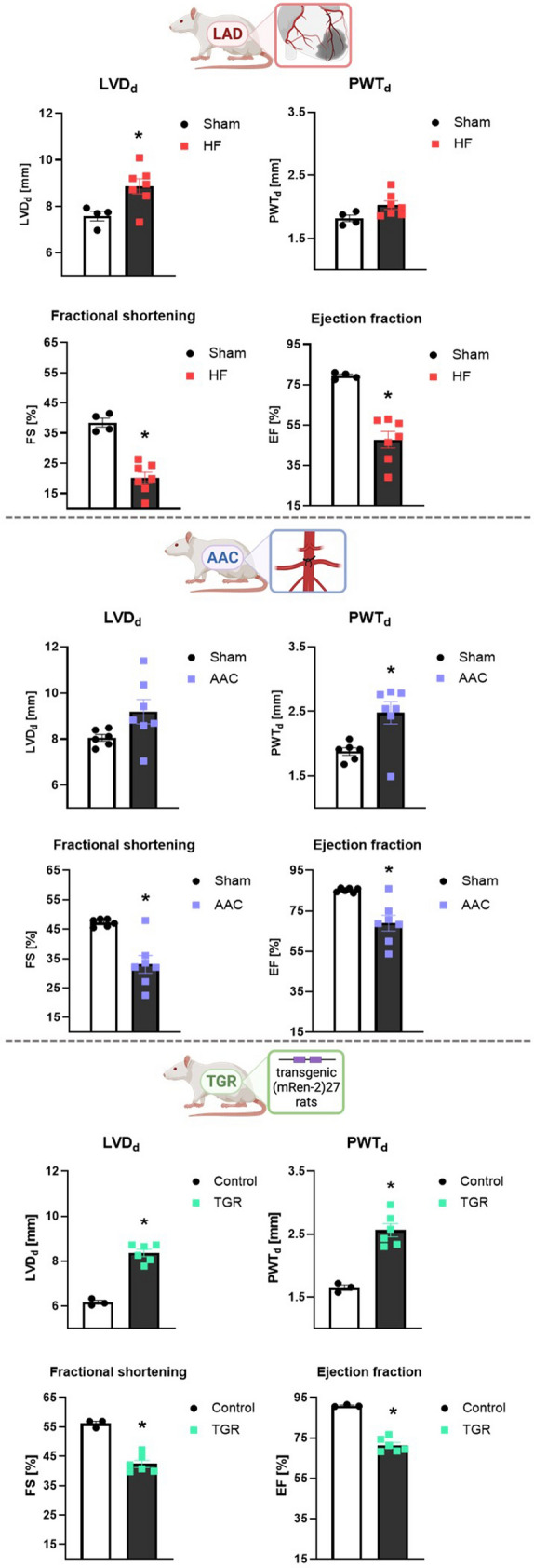
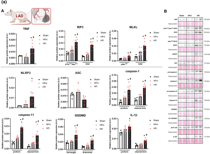
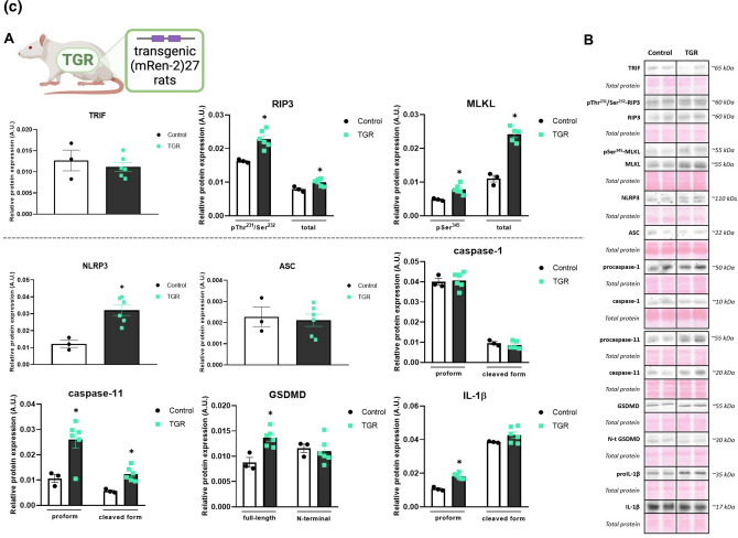
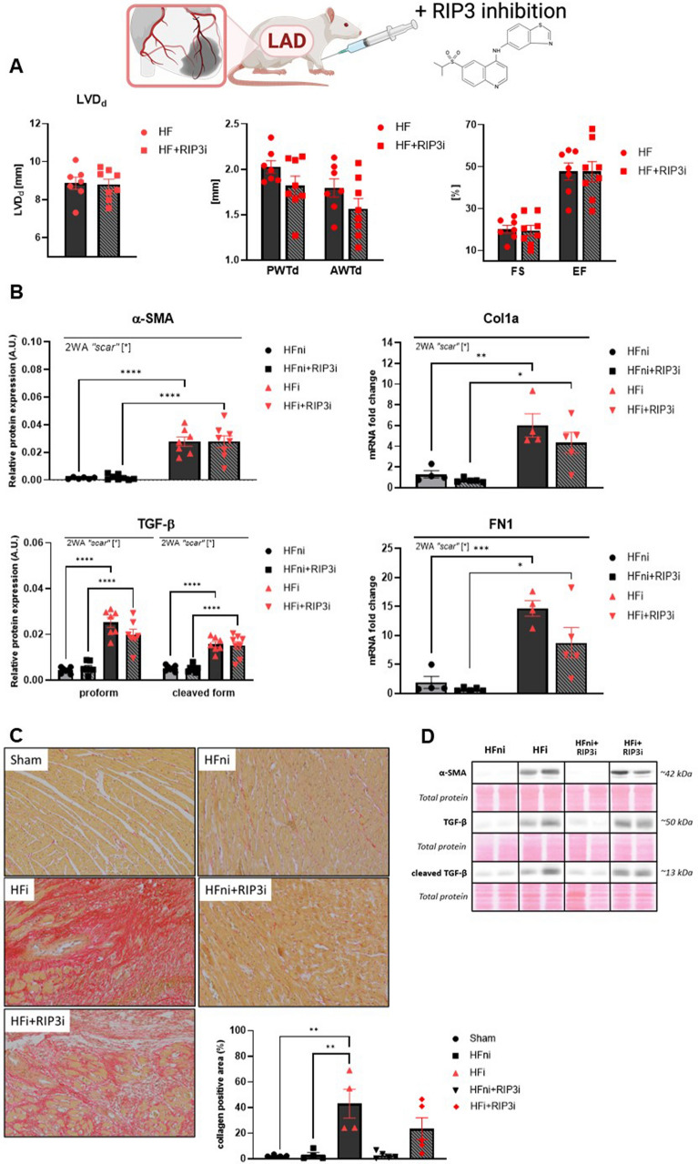
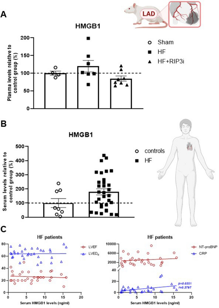
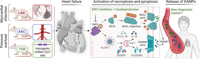
Since cell dying in heart failure (HF) may vary based on the aetiology, we examined the main forms of regulated necrosis, such as necroptosis and pyroptosis, in the hearts damaged due to myocardial infarction (MI) or pressure overload. We also investigated the effects of a drug inhibiting RIP3, a proposed convergent point for both these necrosis-like cell death modes. In rat hearts, left ventricular function, remodelling, pro-cell death, and pro-inflammatory events were investigated, and the pharmacodynamic action of RIP3 inhibitor (GSK'872) was assessed. Regardless of the HF aetiology, the heart cells were dying due to necroptosis, albeit the upstream signals may be different. Pyroptosis was observed only in post-MI HF. The dysregulated miRNAs in post-MI hearts were accompanied by higher levels of a predicted target, HMGB1, its receptors (TLRs), as well as the exacerbation of inflammation likely originating from macrophages. The RIP3 inhibitor suppressed necroptosis, unlike pyroptosis, normalised the dysregulated miRNAs and tended to decrease collagen content and affect macrophage infiltration without affecting cardiac function or structure. The drug also mitigated the local heart inflammation and normalised the higher circulating HMGB1 in rats with post-MI HF. Elevated serum levels of HMGB1 were also detected in HF patients and positively correlated with C-reactive protein, highlighting pro-inflammatory axis. In conclusion, in MI-, but not pressure overload-induced HF, both necroptosis and pyroptosis operate and might underlie HF pathogenesis. The RIP3-targeting pharmacological intervention might protect the heart by preventing pro-death and pro-inflammatory mechanisms, however, additional strategies targeting multiple pro-death pathways may exhibit greater cardioprotection.

Keywords: Heart failure; High mobility group box 1; Inflammation; Necroptosis; Pyroptosis; Receptor-interacting protein kinase 3.


Conflict of interest statement

Declarations. Conflict of interest: The authors declare that they have no conflict of interest.

Figures

Fig. 1
Fig. 1
Echocardiographic assessment of cardiac function in three models of heart failure induced by myocardial infarction or pressure overload, n = 4–7, data are presented as mean ± SEM, unpaired t test or Mann–Whitney test, *P < 0.05
Fig. 2
Fig. 2
A Plasma membrane disrupting mechanisms due to necroptosis and pyroptosis in heart failure (HF) induced by myocardial infarction (MI). A Western blot analysis of MI-induced changes in the expression of necroptotic and pyroptotic proteins (TRIF, pThr231/Ser232-RIP3, RIP3, pSer345-MLKL, MLKL, NLRP3, ASC, (pro)caspase-1, (pro)caspase-11, (N-terminal) GSDMD, (pro)IL-1β) in Sham, non-infarcted HF (HFni) and infarcted HF (HFi) group, n = 4–7, data are presented as mean ± SEM, one-way ANOVA or Kruskal–Wallis test, *P < 0.05 vs Sham, #P < 0.05 vs HFni; B Representative immunoblots and total protein staining. B Plasma membrane disrupting mechanisms due to necroptosis and pyroptosis in heart failure (HF) induced by pressure overload due to abdominal aortic constriction (AAC). A Western blot analysis of AAC-induced changes in the expression of necroptotic and pyroptotic proteins (TRIF, pThr231/Ser232-RIP3, RIP3, pSer345-MLKL, MLKL, NLRP3, ASC, (pro)caspase-1, (pro)caspase-11, (N-terminal) GSDMD, (pro)IL-1β) in Sham and AAC group, n = 5–7, data are presented as mean ± SEM, unpaired t test, *P < 0.05; B Representative immunoblots and total protein staining. C Plasma membrane disrupting mechanisms due to necroptosis and pyroptosis in heart failure (HF) induced by pressure overload in transgenic (mRen-2)27 rats (TGR). A Western blot analysis of TGR-induced changes in the expression of necroptotic and pyroptotic proteins (TRIF, pThr231/Ser232-RIP3, RIP3, pSer345-MLKL, MLKL, NLRP3, ASC, (pro)caspase-1, (pro)caspase-11, (N-terminal) GSDMD, (pro)IL-1β) in Control and TGR group, n = 3–6, data are presented as mean ± SEM, unpaired t test, *P < 0.05; B Representative immunoblots and total protein staining
Fig. 2
Fig. 2
A Plasma membrane disrupting mechanisms due to necroptosis and pyroptosis in heart failure (HF) induced by myocardial infarction (MI). A Western blot analysis of MI-induced changes in the expression of necroptotic and pyroptotic proteins (TRIF, pThr231/Ser232-RIP3, RIP3, pSer345-MLKL, MLKL, NLRP3, ASC, (pro)caspase-1, (pro)caspase-11, (N-terminal) GSDMD, (pro)IL-1β) in Sham, non-infarcted HF (HFni) and infarcted HF (HFi) group, n = 4–7, data are presented as mean ± SEM, one-way ANOVA or Kruskal–Wallis test, *P < 0.05 vs Sham, #P < 0.05 vs HFni; B Representative immunoblots and total protein staining. B Plasma membrane disrupting mechanisms due to necroptosis and pyroptosis in heart failure (HF) induced by pressure overload due to abdominal aortic constriction (AAC). A Western blot analysis of AAC-induced changes in the expression of necroptotic and pyroptotic proteins (TRIF, pThr231/Ser232-RIP3, RIP3, pSer345-MLKL, MLKL, NLRP3, ASC, (pro)caspase-1, (pro)caspase-11, (N-terminal) GSDMD, (pro)IL-1β) in Sham and AAC group, n = 5–7, data are presented as mean ± SEM, unpaired t test, *P < 0.05; B Representative immunoblots and total protein staining. C Plasma membrane disrupting mechanisms due to necroptosis and pyroptosis in heart failure (HF) induced by pressure overload in transgenic (mRen-2)27 rats (TGR). A Western blot analysis of TGR-induced changes in the expression of necroptotic and pyroptotic proteins (TRIF, pThr231/Ser232-RIP3, RIP3, pSer345-MLKL, MLKL, NLRP3, ASC, (pro)caspase-1, (pro)caspase-11, (N-terminal) GSDMD, (pro)IL-1β) in Control and TGR group, n = 3–6, data are presented as mean ± SEM, unpaired t test, *P < 0.05; B Representative immunoblots and total protein staining
Fig. 3
Fig. 3
The effect of RIP3 inhibition on cardiac function and remodelling. A Echocardiographic assessment of cardiac function after 7 days of post-myocardial infarction recovery and the effect of RIP3 inhibition, n = 7–8, data are presented as mean ± SEM, unpaired t test; B Western blot or RT-qPCR analysis of α-SMA, TGF-β, Col1a and FN1 in non-infarcted heart failure (HFni), treated HFni (HFni + GSK´872), infarcted HF (HFi) and treated HFi (HFi + GSK´872) group, n = 4–8, data are presented as mean ± SEM, two-way ANOVA, *P < 0.05, **P < 0.01, ***P < 0.001, ****P < 0.0001; C Representative images showing collagen content and its percentage expression in Sham, non-infarcted HF (HFni), infarcted HF (HFi), treated HFni (HFni + GSK´872) and treated HFi (HFi + GSK´872) group, n = 4–5, data are presented as mean ± SEM, one-way ANOVA, **P < 0.01; D Representative immunoblots and total protein staining
Fig. 4
Fig. 4
The effect of RIP3 inhibition on the plasma membrane disrupting mechanisms. A Schematic illustration of the experimental protocol of post-myocardial infarction (post-MI) heart failure (HF); B Western blot analysis of the necroptotic proteins (TRIF, pThr231/Ser232-RIP3, RIP3, pSer345-MLKL, MLKL, pSer345-MLKL and MLKL oligomeric forms) in non-infarcted HF (HFni), treated HFni (HFni + GSK´872), infarcted HF (HFi) and treated HFi (HFi + GSK´872) group, n = 7–8, data are presented as mean ± SEM, two-way ANOVA, *P < 0.05, **P < 0.01, ****P < 0.0001; C The activity of LDH after 7 days of post-MI recovery in Sham, HF and treated HF (HF + GSK´872) group, n = 4–5, data are presented as mean ± SEM, one-way ANOVA; D Western blot analysis of the pyroptotic proteins (NLRP3, ASC, (pro)caspase-1, (pro)caspase-11, (N-terminal) GSDMD, (pro)IL-1β) in non-infarcted HF (HFni), treated HFni (HFni + GSK´872), infarcted HF (HFi) and treated HFi (HFi + GSK´872) group, n = 7–8, data are presented as mean ± SEM, two-way ANOVA, *P < 0.05, **P < 0.01, ***P < 0.001, ****P < 0.0001; E Identification of macrophages infiltration—representative images showing immunofluorescence staining of F4/80 and its percentage expression and Western blot analysis of Iba1 in Sham, non-infarcted HF (HFni), infarcted HF (HFi), treated HFni (HFni + GSK´872) and treated HFi (HFi + GSK´872) group, n = 4–8, data are presented as mean ± SEM, Kruskal–Wallis test, *P < 0.05; (F) Representative immunoblots and total protein staining
Fig. 5
Fig. 5
The effect of RIP3 inhibition on the alternative mechanisms of necroptosis leading to the plasma membrane rupture. A Western blot analysis of the proteins of alternative necroptosis signallings (pThr286-CaMKIIδ, CaMKIIδ, CypD, pSer637-Drp1, Drp1, PGAM5, pThr183/Tyr185-JNK, JNK, BNIP3, AIF) in non-infarcted heart failure (HFni), treated HFni (HFni + GSK´872), infarcted HF (HFi) and treated HFi (HFi + GSK´872) group, n = 6–8, data are presented as mean ± SEM, two-way ANOVA, *P < 0.05, **P < 0.01, ***P < 0.001, ****P < 0.0001; B Representative immunoblots and total protein staining
Fig. 6
Fig. 6
Analysis of heart failure (HF)-mediated dysregulation of miRNAs and their target proteins including upstream molecules potentially inducing necroptosis and pyroptosis and the effect of RIP3 inhibition. A Venn diagram showing the miRNAs being changed 5- and tenfold in non-infarcted and infarcted HF group compared to Sham group, RT-qPCR analysis of miRNA-140 and miRNA-142 in Sham, non-infarcted HF (HFni), infarcted HF (HFi), treated HFni (HFni + GSK´872) and treated HFi (HFi + GSK´872) group, n = 3–5, data are presented as mean ± SEM, one-way ANOVA, **P < 0.01, ***P < 0.001; ****P < 0.0001; B Western blot or RT-qPCR analysis of the molecules potentially associated with programmed necrosis induction (TLR2, TLR3, TLR4, MyD88, HMGB1, dsHMGB1, frHMGB1, tenascin-C) in Sham, non-infarcted HF (HFni), infarcted HF (HFi), treated HFni (HFni + GSK´872) and treated HFi (HFi + GSK´872) group, n = 4–8, data are presented as mean ± SEM, one-way ANOVA or Kruskal–Wallis test, *P < 0.05, **P < 0.01, ***P < 0.001, ****P < 0.0001; C Representative immunoblots and total protein staining
Fig. 7
Fig. 7
Potential consequences of the plasma membrane rupture—release of HMGB1 into the circulation. A The plasma levels of HMGB1 in Sham rats (n = 4), post-myocardial infarction heart failure (HF) rats (HF) (n = 7) and treated HF rats (HF + GSK´872) (n = 8), data are presented as mean ± SEM, one-way ANOVA; B The serum levels of HMGB1 in human healthy controls (n = 8) and patients with HF (n = 32), data are presented as mean ± SEM, unpaired t test; C Correlation between the serum HMGB1 and LVEF, LVEDd, the serum NT-proBNP and the serum CRP in HF patients (n = 32), Pearson correlation analysis
Fig. 8
Fig. 8
The regulated necrosis-like cell death forms in heart failure due to myocardial infarction or pressure overload, the potential consequences of the plasma membrane rupture and the effect of RIP3 inhibition

References

    1. Adameova A, Goncalvesova E, Szobi A, Dhalla NS (2016) Necroptotic cell death in failing heart: relevance and proposed mechanisms. Heart Fail Rev 21:213–221. 10.1007/s10741-016-9537-8 - PubMed
    1. Badoer E (2022) New Insights Into the Role of Inflammation in the Brain in Heart Failure. Front Physiol 13. 10.3389/FPHYS.2022.837723 - PMC - PubMed
    1. Bai Y, Sun X, Chu Q, Li A, Qin Y, Li Y, Yue E, Wang H, Li GY, Zahra SM, Dong C, Jiang Y (2018) Caspase-1 regulate AngII-induced cardiomyocyte hypertrophy via upregulation of IL-1β. Biosci Rep. 10.1042/BSR20171438 - PMC - PubMed
    1. Bergsbaken T, Fink SL, Cookson BT (2009) Pyroptosis: host cell death and inflammation. Nat Rev Microbiol 7:99–109. 10.1038/nrmicro2070 - PMC - PubMed
    1. Bertheloot D, Latz E, Franklin BS (2021) Necroptosis, pyroptosis and apoptosis: an intricate game of cell death. Cell Mol Immunol 18:1106–1121. 10.1038/S41423-020-00630-3 - PMC - PubMed

MeSH terms

Substances