. 2025 Mar 15;15(1):8991.
doi: 10.1038/s41598-025-92927-4.

Chronic administration of ivabradine improves cardiac Ca handling and function in a rat model of Duchenne muscular dystrophy

Affiliations

Chronic administration of ivabradine improves cardiac Ca handling and function in a rat model of Duchenne muscular dystrophy

Jessica Marksteiner et al. Sci Rep. .

Abstract

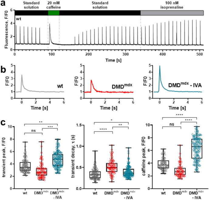
Duchenne muscular dystrophy (DMD), a severe muscle disease caused by mutations in the gene encoding for the intracellular protein dystrophin, is associated with impaired cardiac function and arrhythmias. A causative factor for complications in the dystrophic heart is abnormal calcium (Ca) handling in ventricular cardiomyocytes, and restoration of normal Ca homeostasis has emerged as therapeutic strategy. Here, we used a rodent model of DMD, the dystrophin-deficient DMDmdx rat, to test the following hypothesis: chronic administration of ivabradine (IVA), a drug clinically approved for the treatment of heart failure, improves Ca handling in dystrophic ventricular cardiomyocytes and thereby enhances contractile performance in the dystrophic heart. Intracellular Ca measurements revealed that 4-months administration of IVA to DMDmdx rats significantly improves Ca handling properties in dystrophic ventricular cardiomyocytes. In particular, IVA treatment increased electrically-evoked Ca transients and speeded their decay. This suggested enhanced sarcoplasmic reticulum Ca release and faster removal of Ca from the cytosol. Chronic IVA administration also enhanced the sarcoplasmic reticulum Ca load. Transthoracic echocardiography revealed a significant improvement of cardiac systolic function in IVA-treated DMDmdx rats. Thus, left ventricular ejection fraction and fractional shortening were enhanced, and end-systolic as well as end-diastolic diameters were diminished by the drug. Finally, chronic IVA administration neither significantly attenuated cardiac fibrosis and apoptosis, nor was vascular function improved by the drug. Collectively our findings suggest that long-term IVA administration enhances contractile function in the dystrophic heart by improvement of Ca handling in ventricular cardiomyocytes. Chronic IVA administration may be beneficial for DMD patients.

Keywords: Cardiac function; Cardiomyocyte calcium handling; Duchenne muscular dystrophy; Dystrophic rats; Ivabradine treatment.


Conflict of interest statement

Declarations. Competing interests: The study is reported in accordance with ARRIVE guidelines. No conflicts of interest, financial or otherwise, are declared by the authors.

Figures

Fig. 1
Fig. 1
Ca handling properties of ventricular cardiomyocytes derived from from wt, control DMDmdx, and IVA-treated DMDmdx rats. (a) Original trace example of an intracellular Ca measurement showing the event sequence. Ca transients were first elicited by electrical stimulation at 0.1 Hz frequency. Thereafter, a Ca transient was induced by caffeine application. After caffeine washout, electrical stimulation was started again, and isoprenaline was applied. (b) Representative single electrically-evoked Ca transients of a wt, control DMDmdx, and IVA-treated DMDmdx myocyte at an enlarged time scale in standard extracellular solution. The decay of the electrically-induced Ca signal following the rapid initial rise was fitted with a single exponential function to derive τ-values. (c) left: Comparison of mean Ca peak fluorescence relative to baseline (F/F0) between wt, DMDmdx and IVA-treated DMDmdx myocytes. Each data point represents a single cell, and values are expressed as median, interquartile range, and minimum/maximum. [107 cells for wt (5 animals); 97 cells for DMDmdx (6 animals) and 177 cells for IVA-treated DMDmdx (6 animals) myocytes]. (c) middle: Comparison of the Ca transient decay kinetics in wt, DMDmdx and IVA-treated DMDmdx myocytes. (c) right: Comparison of mean Ca peak fluorescence, elicited by caffeine application, relative to baseline (F/F0) between wt, DMDmdx and IVA-treated DMDmdx myocytes [82 cells for wt (5 animals); 81 cells for DMDmdx (5 animals) and 167 cells for IVA-treated DMDmdx (6 animals) myocytes]. *p < 0.05, **p < 0.01, ***p < 0,001, ****p < 0,0001; ns, not significant.
Fig. 2
Fig. 2
The expression of Ca handling proteins in left ventricular tissue samples. Representative western blots showing SERCA2 (a), annexin 6A (b) phospholamban (c, top), phospho (Ser16/Thr17)-phospholamban (c, bottom) and sarcolipin (d, monomer and multimer formation) protein levels in left ventricular tissue, which was obtained from wt, DMDmdx and IVA-treated DMDmdx rats. This figure also shows the same lanes after membranes were re-probed for GAPDH or vinculin to demonstrate equal protein loading in each lane. Semi-quantitative analysis of SERCA2 (e), annexin 6A (f) phospholamban (g) and phospho (Ser16/Thr17)- phospholamban (h), phospho (Ser16/Thr17)- phospholamban/phospholamban (i) and sarcolipin (j–l) in left ventricular tissue. Data represent means ± SEM; n = 8 for wt, n = 6 for DMDmdx, and n = 6 for IVA-treated DMDmdx. Statistical comparisons were carried out using a one-way ANOVA and post-hoc Tukey’s multiple comparison test, **p < 0.01. DMD Duchenne Muscular Dystrophy, GAPDH glyceraldehyde 3-phosphate dehydrogenase, IVA ivabradine, PLN phospholamban, p-PLN phospho (Ser16/Thr17)-phospholamban, SERCA2 sarcoplasmic/endoplasmic reticulum Ca ATPase 2, ns not significant, wt wild-type.
Fig. 3
Fig. 3
Heart function and dimensions assessed by transthoracic echocardiography. Comparison of echocardiography parameters between wt, DMDmdx and IVA-treated DMDmdx rats. All parameters were measured for at least three successive cardiac cycles. LVEF: left ventricular ejection fraction (a); LVFS: left ventricular fractional shortening (b); LVESD: Left ventricular end-systolic diameter (c); LVEDD: left ventricular end-diastolic diameter (d). Data represent means ± SEM; n = 8 wt rats; n = 13 DMDmdx rats, and n = 13 IVA-treated DMDmdx rats. Statistical comparisons were carried out using a one-way ANOVA and post-hoc Tukey’s multiple comparison test, *p < 0.05, **p < 0.01 and ***p < 0.001. DMD: Duchenne Muscular Dystrophy, IVA: ivabradine; ns: not significant, and wt: wild-type.
Fig. 4
Fig. 4
The impact of chronic ivabradine administration on cardiac fibrosis and apoptosis. (a) Representative pictures of haematoxylin & eosin (HE)-stained cardiac sections, and representative images of myocardial fibrosis stained with the Masson trichrome method (MG). Cardiac sections derived from untreated control DMDmdx rats (left) and IVA-treated DMDmdx rats (right) are compared. (b) Representative photographs of TUNEL staining in cardiac sections of untreated DMDmdx (left) and IVA-treated DMDmdx (right) rats. This assay was performed to assess the degree and distribution of apoptotic cells in normal and fibrotic/remodelled heart tissue. Green dots represent apoptotic cells, resulting from the overlay of TUNEL-stained and DAPI-counterstained nuclei. *Areas with inflammatory cells which are TUNEL-negative. Blue dots represent non-apoptotic cells stained with DAPI only. Scale bar = 50 µm (200 × magnification). DAPI 4′,6-diamidino-2-phenylindole, DMD Duchenne Muscular Dystrophy, IVA ivabradine.
Fig. 5
Fig. 5
Vascular function analysis. Endothelium-dependent (a) and endothelium-independent (b) relaxation curves from isolated rings of abdominal aortas from wt (empty circles), DMDmdx (black circles), and IVA-treated DMDmdx (grey circles) rats in response to increasing concentrations of acetylcholine (ACh) and sodium nitroprusside (SNP), respectively. Data represent means ± SEM; n = 17 for wt, n = 15 for DMDmdx, and n = 15 for IVA + DMDmdx aorta segments. Statistical comparisons were carried out using a repeated measure-ANOVA and post-hoc Tukey’s multiple comparison test, **p < 0.01 and ***p < 0.001 vs wt. DMD Duchenne Muscular Dystrophy, IVA ivabradine, wt wild-type.

References

    1. Rushworth, G. F., Lambrakis, P. & Leslie, S. J. Ivabradine: A new rate-limiting therapy for coronary artery disease and heart failure. Ther. Adv. drug Saf.2, 19–28 (2011). - PMC - PubMed
    1. Scicchitano, P. et al. Ivabradine, coronary artery disease, and heart failure: Beyond rhythm control. Drug Des. Devel. Ther.8, 689–700 (2014). - PMC - PubMed
    1. Koruth, J. S., Lala, A., Pinney, S., Reddy, V. Y. & Dukkipati, S. R. The clinical use of ivabradine. J. Am. Coll. Cardiol.70, 1777–1784 (2017). - PubMed
    1. Yang, J. et al. The effect of ivabradine therapy on dilated cardiomyopathy patients with congestive heart failure: A systematic review and meta-analysis. Front. Cardiovasc. Med.10, 1149351 (2023). - PMC - PubMed
    1. Finsterer, J., Stöllberger, C. & Berger, E. Beneficial effect of ivabradine in dilated cardiomyopathy from Becker muscular dystrophy. Herz37, 702–705 (2012). - PubMed

MeSH terms

LinkOut - more resources