. 2025 Mar 15;22(1):86.
doi: 10.1186/s12974-025-03406-6.

Targeted activation of ErbB4 receptor ameliorates neuronal deficits and neuroinflammation in a food-borne polystyrene microplastic exposed mouse model

Affiliations

Targeted activation of ErbB4 receptor ameliorates neuronal deficits and neuroinflammation in a food-borne polystyrene microplastic exposed mouse model

Chong Liu et al. J Neuroinflammation. .

Abstract

The impact of polystyrene microplastics (PS-MPs) on the nervous system has been documented in the literature. Numerous studies have demonstrated that the activation of the epidermal growth factor receptor 4 (ErbB4) is crucial in neuronal injury and regeneration processes. This study investigated the role of targeted activation of ErbB4 receptor through a small molecule agonist, 4-bromo-1-hydroxy-2-naphthoic acid (C11H7BrO3, E4A), in mitigating PS-MPs-induced neuronal injury. The findings revealed that targeted activation of ErbB4 receptor significantly ameliorated cognitive behavioral deficits in mice exposed to PS-MPs. Furthermore, E4A treatment upregulated the expression of dedicator of cytokinesis 3 (DOCK3) and Sirtuin 3 (SIRT3) and mitigated mitochondrial and synaptic dysfunction within the hippocampus of PS-MPs-exposed mice. E4A also diminished the activation of the TLR4-NF-κB-NLRP3 signaling pathway, consequently reducing neuroinflammation. In vitro experiments demonstrated that E4A partially alleviated PS-MPs-induced hippocampal neuronal injury and its effects on microglial inflammation. In conclusion, the findings of this study indicate that targeted activation of ErbB4 receptor may mitigate neuronal damage and subsequent neuroinflammation, thereby alleviating hippocampal neuronal injury induced by PS-MPs exposure and ameliorating cognitive dysfunction. These results offer valuable insights for the development of potential therapeutic strategies.

Keywords: Cognitive dysfunction; ErbB4 receptor; Mitochondrial damage; Neuroinflammation; Neuronal damage; PS-MPs; Small molecule.


Conflict of interest statement

Declarations. Ethical approval: All procedures were conducted according to the Ethics Committee of Jiangnan University Wuxi Medical College. Competing interests: The authors declare no competing interests.

Figures

Fig. 1
Fig. 1
ErbB4 small molecule agonist ameliorates cognitive behavioral impairments in mice exposed to PS-MPs. (A) Representative traces in open field experiment. (B) The statistical analysis related to open field test. (C) Representative heat map in Y maze exploration experiment, with arm A denoting the initial arm. (D) The rate of spontaneous alternation in Y maze experiment. (E) Representative traces from the new object recognition (NOR) experiment. (F) Statistical analysis related to the NOR experiment. (G) Representative swimming traces from the Morris water maze (MWM) exploration experiment. (H) The statistical analysis of MWM exploration test. Statistical analysis was conducted using one-way analysis of variance (ANOVA) followed by Tukey’s post hoc test for multiple comparisons, and the data are presented as means ± standard error of the mean (SEM). Statistical significance is indicated by *p < 0.05, **p < 0.01, and ***p < 0.001, with n = 8 per group
Fig. 2
Fig. 2
ErbB4 small molecule agonist can improve the dendritic spine injury of hippocampal neurons in mice exposed to PS-MPs. (A) Representative images of neuronal dendritic segments. scale bar, 10 μm. (B) Total dendritic spine density of hippocampal neurons. (C) The density of mushroom spines in hippocampal neurons. Statistical analysis was conducted using one-way ANOVA followed by Tukey’s post hoc test for multiple comparisons.The results were presented as means ± SEM. Statistical significance is indicated by *p < 0.05, **p < 0.01, and ***p < 0.001, with n = 4 per group
Fig. 3
Fig. 3
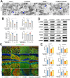
ErbB4 small molecule agonist can improve synaptic dysfunction of hippocampal neurons in mice exposed to PS-MPs. (A) The ultrastructural analysis of hippocampal neuron synapses was performed using transmission electron microscopy (TEM). (B) Statistical analyses were conducted on the synaptic data of hippocampal neurons. (C) Immunofluorescence was employed to detect the levels of synaptophysin (SYP) and β3-tubulin. (D) Western blot analysis was utilized to assess the protein levels of synapse-associated proteins, including DOCK3, p-CREB, CREB, SYP, GAP43, PSD95, and SYT1 in hippocampal neurons. (E) Statistical analyses of these protein levels were performed using one-way ANOVA with Tukey’s multiple comparison test. The synaptic structure was examined at a magnification of 10,000×, with a scale bar of 1 μm. Results are expressed as means ± SEM. Immunofluorescence labeling was as follows: SYP in red and β3-tubulin in green, with a scale bar of 20 μm. Statistical significance was indicated by *p < 0.05, **p < 0.01, and ***p < 0.001, with a sample size of n = 4 per group
Fig. 4
Fig. 4
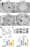
ErbB4 small molecule agonist can improve mitochondrial damage in hippocampal neurons of mice exposed to PS-MPs. (A) The ultrastructural integrity of hippocampal neuronal mitochondria was evaluated using transmission electron microscopy (TEM). (B) Statistical analyses were performed on data pertaining to the hippocampal neuronal mitochondria. (C) The expression levels of mitochondria-associated proteins, including phosphorylated mTOR (p-mTOR), mTOR, and SIRT3, in the hippocampus of mice were quantified via western blot analysis. (D) Statistical analyses of these protein expression levels were conducted using one-way ANOVA followed by Tukey’s post hoc test for multiple comparisons. The original magnification for mitochondrial imaging was 2000×, with a scale bar of 5 μm. Data are presented as means ± SEM. Statistical significance was indicated by *p < 0.05, **p < 0.01, and ***p < 0.001, with a sample size of n = 4 per group
Fig. 5
Fig. 5
ErbB4 small molecule agonist enhances the ErbB4 signaling pathway in hippocampus of mice exposed to PS-MPs. (A) Immunofluorescence was used to detect levels of phosphorylated ErbB4 (p-ErbB4) and β3-tubulin. (B) Protein levels associated with the ErbB4 signaling pathway in the hippocampus of mice were assessed through western blot analysis. (C) A statistical analysis was performed on the data derived from protein levels, employing one-way ANOVA with Tukey’s multiple comparison tests. The p-ErbB4 is represented in red, while β3-tubulin is depicted in green, with a scale bar of 20 μm. The results are expressed as means ± SEM, with significance levels indicated by *p < 0.05, **p < 0.01, and ***p < 0.001. Each experimental group comprised n = 4 samples
Fig. 6
Fig. 6
ErbB4 small molecule agonist can improve hippocampal neuroinflammation in mice exposed to PS-MPs via the TLR4/NLRP3 pathway. (A) Levels of IBA1 were detected through immunofluorescence staining. (B) Levels of GFAP were detected through immunofluorescence staining. (C) Western blot analysis was utilized to detect protein levels associated with the TLR4/NLRP3 signaling pathway in the hippocampus of mice. (D) Statistical chart was generated to display the protein levels of TLR4/NLRP3. GFAP and IBA1 are both indicated in red, with a scale bar of 20 μm. Statistical analysis was conducted using one-way ANOVA followed by Tukey’s post hoc test for multiple comparisons, and the data are reported as means ± SEM, with significance levels denoted as *p < 0.05, **p < 0.01, and ***p < 0.001. Each group comprised n = 4 samples
Fig. 7
Fig. 7
The ErbB4 small molecule agonist ameliorates hippocampal neuron damage under pathological conditions induced by PS-MPs in vitro via the ErbB4 signaling pathway, and concurrently attenuates microglial activation through the TLR4/NLRP3 signaling pathway. (A-D) Following exposure to PS-MPs at a concentration of 100 µg/mL for 24 h, hippocampal neurons were subsequently treated with C11H7BrO3 at a concentration of 10 nM for an additional 24 h. (A, C) Protein expression levels associated with the ErbB4 signaling pathway, mitochondrial function, apoptosis, and synaptic integrity in hippocampal neurons were quantified using Western blot analysis. (B, D) Statistical representations of these protein expression levels were depicted in graphical form. (E) Oxidative stress markers in hippocampal neurons, including total superoxide dismutase (T-SOD), glutathione (GSH), glutathione peroxidase (GSH-Px), and malondialdehyde (MDA) levels, were evaluated. (F) Intracellular ATP levels in hippocampal neurons were also measured. (G-H) Microglial cells were co-cultured with supernatants derived from treated hippocampal neurons for 24 h. (G) Protein expression levels related to the TLR4/NLRP3 signaling pathway in microglial cells were assessed via Western blot analysis. (H) Statistical representations of these protein expression levels were also depicted in graphical form. Statistical analysis was conducted using one-way ANOVA followed by Tukey’s post hoc test for multiple comparisons, and data are presented as means ± SEM. Significance levels were denoted as *p < 0.05, **p < 0.01, ***p < 0.001. A minimum sample size of n = 4–6 per group was utilized
Fig. 8
Fig. 8
The schematic diagram illustrates the impact of targeted activation of ErbB4 receptor on hippocampal neuronal damage, neuroinflammation, and cognitive functions in mice exposed to PS-MPs. The small molecule ErbB4 receptor agonist (E4A) binds to the ErbB4 receptor, initiating its downstream signaling pathway. This activation leads to the upregulation of SIRT3 protein expression, which enhances mitochondrial redox homeostasis, increases ATP production, and ameliorates mitochondrial dysfunction. Additionally, there is an upregulation of synaptic-related proteins, which contributes to the repair of synaptic damage and enhancement of synaptic function. As hippocampal neuronal function improves, the activation of the TLR4 receptor by pro-inflammatory factors in microglia is reduced, resulting in diminished phosphorylation of NF-κB and decreased activation of the NLRP3 inflammasome towards Caspase-1. Consequently, there is a reduction in the conversion of the IL-1β precursor to its mature form, thereby alleviating hippocampal neuroinflammation and ultimately enhancing the learning and cognitive functions of PS-MPs-exposed mice. The diagram was created with MedPeer (www.medpeer.cn)

References

    1. Zhao B, Rehati P, Yang Z, Cai Z, Guo C, Li Y. The potential toxicity of microplastics on human health. Sci Total Environ. 2024;912:168946. 10.1016/j.scitotenv.2023.168946. - PubMed
    1. Allouzi MMA, Tang DYY, Chew KW, Rinklebe J, Bolan N, Allouzi SMA, et al. Micro (nano) plastic pollution: the ecological influence on soil-plant system and human health. Sci Total Environ. 2021;788:147815. 10.1016/j.scitotenv.2021.147815. - PubMed
    1. Cox KD, Covernton GA, Davies HL, Dower JF, Juanes F, Dudas SE. Human consumption of microplastics. Environ Sci Technol. 2019;53(12):7068–74. 10.1021/acs.est.9b01517. - PubMed
    1. Deng Y, Zhang Y, Lemos B, Ren H. Tissue accumulation of microplastics in mice and biomarker responses suggest widespread health risks of exposure. Sci Rep. 2017;7:46687. 10.1038/srep46687. - PMC - PubMed
    1. Senathirajah K, Attwood S, Bhagwat G, Carbery M, Wilson S, Palanisami T. Estimation of the mass of microplastics ingested - A pivotal first step towards human health risk assessment. J Hazard Mater 404(Pt B). 2021;124004. 10.1016/j.jhazmat.2020.124004. - PubMed

LinkOut - more resources