

Optogenetic stimulation of cell bodies versus axonal terminals generate comparable activity and functional connectivity patterns in the brain
- PMID: 40090667
- PMCID: PMC12165442
- DOI: 10.1016/j.brs.2025.03.006
Optogenetic stimulation of cell bodies versus axonal terminals generate comparable activity and functional connectivity patterns in the brain
Abstract
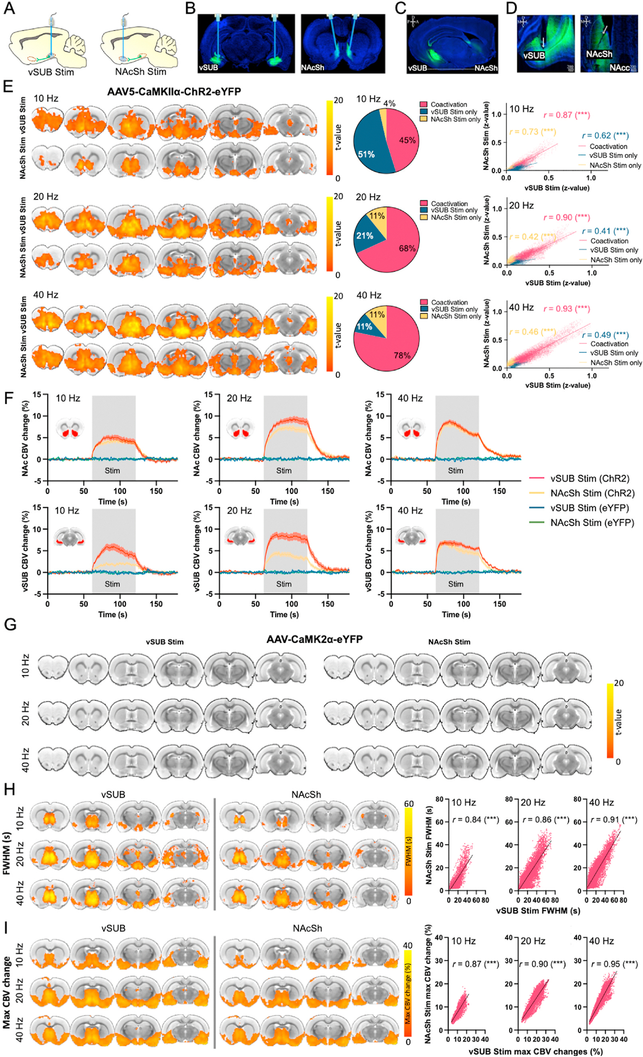
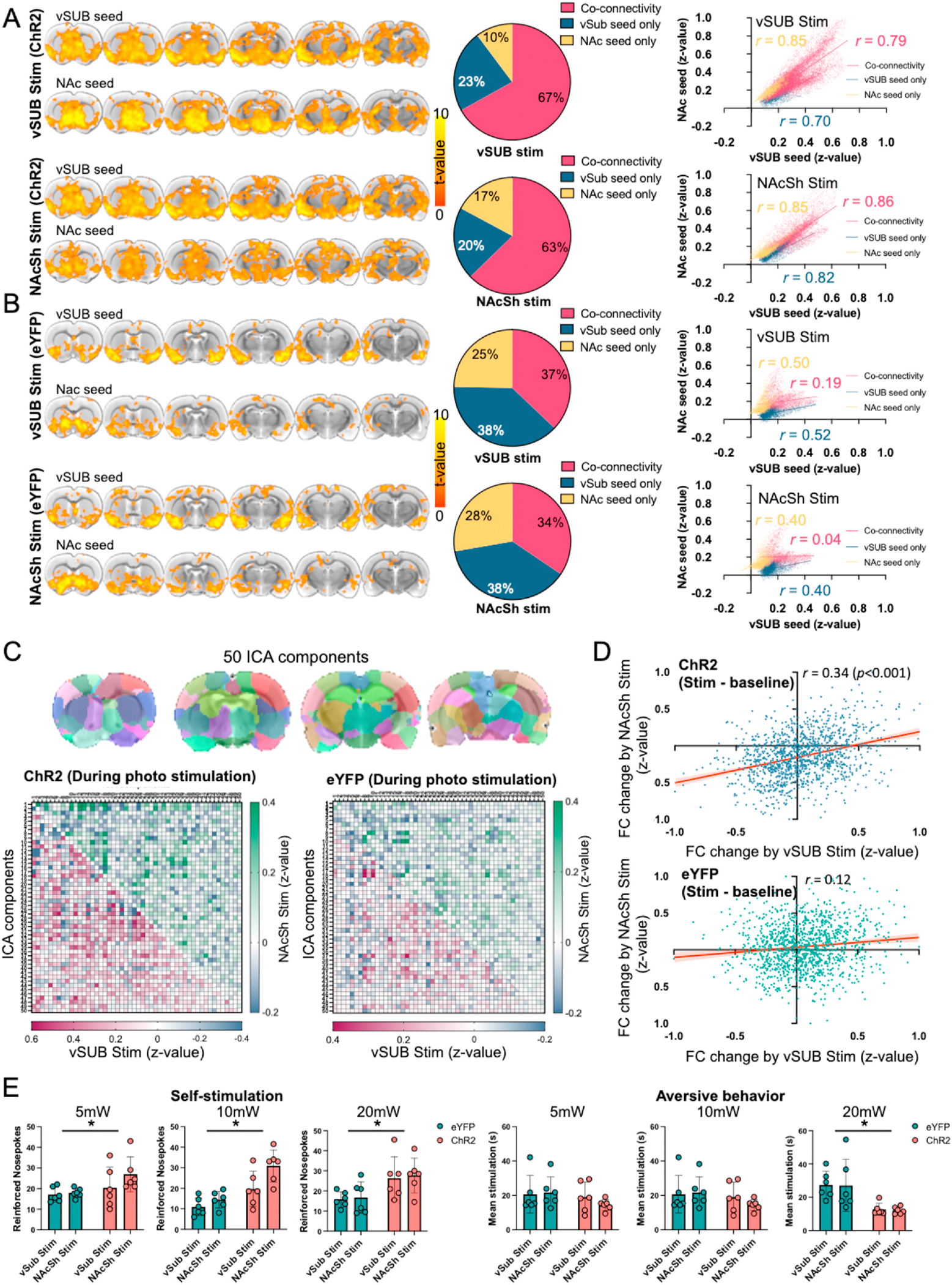
Optogenetic techniques are often employed to dissect neural pathways with presumed specificity for targeted projections. In this study, we used optogenetic fMRI to investigate the effective landscape of stimulating the cell bodies versus one of its projection terminals. Specifically, we selected a long-range unidirectional projection from the ventral subiculum (vSUB) to the nucleus accumbens shell (NAcSh) and placed two stimulating fibers-one at the vSUB cell bodies and the other at the vSUB terminals in the NAcSh. Contrary to the conventional view that terminal stimulation confines activity to the feedforward stimulated pathway, our findings reveal that terminal stimulation induces brain activity and connectivity patterns remarkably similar to those of vSUB cell body stimulation. This observation suggests that the specificity of optogenetic terminal stimulation may induce antidromic activation, leading to broader network involvement than previously acknowledged.
Keywords: Antidromic activation; Functional connectivity; Nucleus accumbens shell (NAcSh); Optogenetics; Ventral subiculum (vSUB); fMRI.
Copyright © 2025 The Authors. Published by Elsevier Inc. All rights reserved.
Conflict of interest statement
Declaration of competing interest The authors declare that they have no known competing financial interests or personal relationships that could have appeared to influence the work reported in this paper.
Figures
Similar articles
-
Ventral subiculum promotes wakefulness through several pathways in male mice.Neuropsychopharmacology. 2024 Aug;49(9):1468-1480. doi: 10.1038/s41386-024-01875-6. Epub 2024 May 11. Neuropsychopharmacology. 2024. PMID: 38734818 Free PMC article.
-
Ventral Subiculum Inputs to Nucleus Accumbens Medial Shell Preferentially Innervate D2R Medium Spiny Neurons and Contain Calcium Permeable AMPARs.J Neurosci. 2023 Feb 15;43(7):1166-1177. doi: 10.1523/JNEUROSCI.1907-22.2022. Epub 2023 Jan 6. J Neurosci. 2023. PMID: 36609456 Free PMC article.
-
Enhancing excitatory projections from the ventral subiculum to the nucleus accumbens shell contribute to the MK-801-induced impairment of prepulse inhibition.Neurosci Lett. 2020 Jul 13;731:135024. doi: 10.1016/j.neulet.2020.135024. Epub 2020 May 4. Neurosci Lett. 2020. PMID: 32380142
-
A role for the subiculum in the brain motivation/reward circuitry.Behav Brain Res. 2006 Nov 11;174(2):225-31. doi: 10.1016/j.bbr.2006.05.036. Epub 2006 Jul 25. Behav Brain Res. 2006. PMID: 16870273 Review.
-
Informing brain connectivity with optogenetic functional magnetic resonance imaging.Neuroimage. 2012 Oct 1;62(4):2244-9. doi: 10.1016/j.neuroimage.2012.01.116. Epub 2012 Feb 3. Neuroimage. 2012. PMID: 22326987 Review.
References
MeSH terms
Grants and funding
LinkOut - more resources
Full Text Sources
