. 2025 Apr 11;136(8):830-844.
doi: 10.1161/CIRCRESAHA.124.325848. Epub 2025 Mar 17.

Overexpression of Wild-Type TMEM43 Improves Cardiac Function in Arrhythmogenic Right Ventricular Cardiomyopathy Type 5

Affiliations

Overexpression of Wild-Type TMEM43 Improves Cardiac Function in Arrhythmogenic Right Ventricular Cardiomyopathy Type 5

Laura Lalaguna et al. Circ Res. .

Abstract

Background: Arrhythmogenic right ventricular cardiomyopathy type 5 (ARVC5) is the most aggressive type of ARVC, caused by a fully penetrant missense mutation (p.S358L) in TMEM43 (transmembrane protein 43). Pathologically, the disease is characterized by dilation of the cardiac chambers and fibrofatty replacement of the myocardium, which results in heart failure and sudden cardiac death. Current therapeutic options are limited, and no specific therapies targeting the primary cause of the disease have been proposed.

Methods: We investigated whether overexpression of wild-type (WT) TMEM43 could overcome the detrimental effects of the mutant form. We used transgenic mouse models overexpressing either WT or mutant (S358L) TMEM43 to generate a double transgenic mouse line overexpressing both forms of the protein. In addition, we explored if systemic delivery of a codon-optimized self-complementary adeno-associated virus bearing WT-TMEM43 could improve disease progression assessed by ECG and echocardiography.

Results: Double transgenic mice overexpressing both WT and mutant TMEM43 forms showed delayed ARVC5 onset, improved cardiac contraction, and reduced ECG abnormalities compared with mice expressing S358L-TMEM43. In addition, cardiomyocyte death and myocardial fibrosis were reduced, with an overall increase in survival. Finally, we demonstrated that a single systemic administration of an adeno-associated virus carrying codon-optimized WT-TMEM43 prevents ventricular dysfunction and ECG abnormalities induced by S358L-TMEM43.

Conclusions: Overexpression of WT-TMEM43 improves the pathological phenotype in a mouse model of ARVC5. Adeno-associated virus-mediated delivery of WT-TMEM43 offers a promising and specific therapy for patients suffering from this highly lethal disease.

Keywords: arrhythmogenic right ventricular dysplasia; cardiomyopathies; mice, transgenic; myocardium.


Conflict of interest statement

The authors would like to acknowledge that this research has led to the filing of a pending patent application. Inventors: B.J. Byrne, M. Corti, E. Lara-Pezzi, L. Lalaguna, and P. García-Pavía. Application number: EP25382006.2. Title: Method for improving cardiac function in arrhythmogenic right ventricular cardiomyopathy type 5. Priority date: 08/01/2025. Applicants: Centro Nacional de Investigaciones Cardiovasculares Carlos III (F.S.P.), Fundación para la Investigación Biomédica del Hospital Universitario Puerta de Hierro Majadahonda, University of Florida Research Foundation, Incorporated.

Figures

Figure 1.
Figure 1.
Wild-type (WT) TMEM43 (transmembrane protein 43) overexpression improves systolic function and life span in arrhythmogenic right ventricular cardiomyopathy type 5 (ARVC5) mice. A, Anti-GFP (green fluorescent protein) co-immunoprecipitation in protein extracts from P19 cells transfected with GFP- and HA-tagged WT (blue) or S358L (MUT, red) TMEM43. Input (In), supernatant (Sn), and output (Out) from the IP (immunoprecipitation) were analyzed by Western blot. TMEM43 was detected by Western blot with antibodies against GFP and HA. B, Survival rate of WT mice (n=22), mice overexpressing either WT-TMEM43 (TMEM43wt, n=20) or S358L-TMEM43 (TMEM43mut, n=35), and double-transgenic mice (n=35). The indicated P value was obtained with a log-rank test. C through F, Left ventricular ejection fraction (LVEF, C), left ventricle end-diastolic volume (LVEDV, D), left ventricle posterior wall thickness in diastole (LVPWd, E), and tricuspid annular plane systolic excursion (TAPSE, F) measured by echocardiography at 5, 16, and 24 weeks of age. n=16 to 36, each group. G, Ratio of heart weight to tibial length (HW/TL) determined at 5, 16, and 24 weeks. n=13 to 31, each group. Data are shown as mean±SD. Two-way ANOVA followed by Bonferroni posttest (C and G) or multiple Mann-Whitney U tests followed by Bonferroni-Dunn posttest (D, E, and F) were used. Since there is no significant difference between WT and TMEM43wt mice, statistical comparisons between TMEM43wt and TMEM43mut or double transgenic mice are omitted to enhance the clarity of the plots. HA indicates hemagglutinin; and IB, immunoblot.
Figure 2.
Figure 2.
Wild-type (WT) TMEM43 (transmembrane protein 43) overexpression reduces ECG abnormalities in arrhythmogenic right ventricular cardiomyopathy type 5 (ARVC5) mice. A, Surface ECG traces and magnifications from lead II in 16-week-old mice. B through D, P-wave duration (B) and QRS duration and amplitude (C and D) were measured from lead II surface electrocardiograms. n=16 to 32, each group. Data are shown as mean±SD. Multiple Mann-Whitney U tests were used, followed by Bonferroni-Dunn posttest. Since there is no significant difference between WT and TMEM43wt mice, statistical comparisons between TMEM43wt and TMEM43mut or double transgenic mice are omitted to enhance the clarity of the plots.
Figure 3.
Figure 3.
Wild-type (WT) TMEM43 (transmembrane protein 43) overexpression results in reduced cardiomyocyte death and downregulation of fibrosis-related genes. A, Serum troponin I (TnI) levels were measured by ELISA in mice at 5, 10, 16, and 24 weeks of age. n=6 to 21, each group. B through F, mRNA expression levels of LOX (lysyl oxidase, B), POSTN (periostin, C), COL1A1 (collagen type I alpha 1 chain, D), COL3A1 (collagen type III alpha 1 chain, E), and ACTA1 (alpha actin 1, F) were determined by quantitative reverse-transcribed polymerase chain reaction in 16-week-old mouse hearts. n=8, each group. Data are shown as mean±SD. Multiple Mann-Whitney U tests followed by Bonferroni-Dunn posttest (A) or 1-way ANOVA followed by Bonferroni posttest (B through F) were used. Since there is no significant difference between WT and TMEM43wt mice, statistical comparisons between TMEM43wt and TMEM43mut or double transgenic (DT) mice are omitted to enhance the clarity of the plots.
Figure 4.
Figure 4.
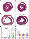
Wild-type (WT) TMEM43 (transmembrane protein 43) overexpression reduces myocardial fibrosis in arrhythmogenic right ventricular cardiomyopathy type 5 (ARVC5) mice. A through D, Collagen content in myocardial cross-sections from WT (A), TMEM43wt (B), TMEM43mut (C), and double transgenic (D) mice was analyzed at 24 weeks of age by Masson trichrome staining; scale bar, 500 µm. E, Quantification of fibrotic tissue from Masson trichrome staining. n=6 to 8, each group. F, Brillouin frequency shift was obtained from myocardial sections of 24-week-old mice. n=4 to 11, each group. Data are shown as mean±SD. Multiple Mann-Whitney U tests followed by Bonferroni-Dunn posttest were used. Since there is no significant difference between WT and TMEM43wt mice, statistical comparisons between TMEM43wt and TMEM43mut or double transgenic mice are omitted to enhance the clarity of the plots. IVS indicates interventricular septum; LV, left ventricle; and RV, right ventricle.
Figure 5.
Figure 5.
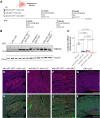
Delivery of codon-optimized wild-type (WT)-TMEM43 (transmembrane protein 43) using an adeno-associated viral vector (AAV) shows high expression in the myocardium. A, Experimental design of self-complementary AAV administration and outcome analyses. A total viral load of 5e10 viral particles (VP) was administered into postnatal day 1 (P1) mice through the temporal vein as follows: AAV carrying TMEM43WT-OPT (AAV-WT-OPT)+AAV carrying luciferase (AAV-LUC; 2.5e10 VP each), AAV carrying S358L-TMEM43 (AAV-MUT)+AAV-LUC (2.5e10 VP each), or AAV-WT-OPT+AAV-MUT (2.5e10 VP each). B, TMEM43 expression was analyzed by western blot. n=6, each group. C, Luciferase mRNA expression levels were determined by quantitative reverse-transcribed polymerase chain reaction in 16-week-old mice hearts after intravenous delivery of AAV-LUC (5e10 VP/neonate), AAV-WT-OPT+AAV-LUC (2.5e10 VP/neonate each), AAV-MUT+AAV-LUC (2.5e10 VP/neonate each), or AAV-WT-OPT+AAV-LUC (2.5e10 VP/neonate each). n=5 to 9, each group. D through G, TMEM43 immunofluorescence analysis, and wheat germ agglutinin (WGA) co-stains in myocardial sections of 16-week-old mice after neonatal injection of AAVs; scale bar, 200 µm. Data are shown as mean±SD (C). One-way ANOVA was used, followed by Bonferroni posttest.
Figure 6.
Figure 6.
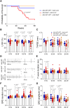
Early codon-optimized wild-type TMEM43 AAV vector (AAV-WT-OPT) administration improves life span, systolic function, and cardiac conduction defects. A, Survival rate of AAV-WT-OPT+AAV vector expressing luciferase (AAV-LUC; n=12), AAV vector expressing S358L-TMEM43 (AAV-MUT)+AAV-LUC (n=19), and AAV-WT-OPT+AAV-MUT (n=13). The indicated P value was obtained with a log-rank test. B and C, Left ventricular ejection fraction (LVEF, B) and tricuspid annular plane systolic excursion (TAPSE, C) at 5, 10, and 16 weeks after AAV administration. D through G, Surface ECG at 16 weeks after AAV injection (D). P-wave duration (E) and QRS duration and amplitude (F and G) at 5, 10, 16, and 24 weeks after AAV injection. n=8 to 28, each group. Two-way ANOVA followed by Bonferroni posttest (B and C) or multiple Mann-Whitney U tests followed by Bonferroni-Dunn posttest (E, F, and G) were used.
Figure 7.
Figure 7.
Codon-optimized wild-type TMEM43 AAV vector (AAV-WT-OPT) reduces cardiomyocyte necrosis and expression of fibrosis-related genes in mice. A, Serum troponin I (TnI) by ELISA in mice at 5, 10, and 16 weeks of age. n=6, each group. B through F, mRNA expression levels of LOX (lysyl oxidase, B), POSTN (periostin, C), COL1A1 (collagen type I alpha 1 chain, D), COL3A1 (collagen type III alpha 1 chain, E) and ACTA1 (alpha actin 1, F) quantified by quantitative reverse-transcribed polymerase chain reaction in 16 weeks old mouse hearts. n=7 to 9, each group. Data are shown as mean±SD. Multiple Mann-Whitney U tests followed by Bonferroni-Dunn posttest (A) or 1-way ANOVA (B through F) were used, followed by Bonferroni posttest. AAV-LUC indicates AAV vector expressing luciferase; and AAV-MUT, AAV vector expressing S358L-TMEM43.
Figure 8.
Figure 8.
Codon-optimized wild-type TMEM43 AAV vector (AAV-WT-OPT) treatment in symptomatic arrhythmogenic right ventricular cardiomyopathy type 5 (ARVC5) adult mice improves systolic function and reduces arrhythmia susceptibility. A, Experimental design of symptomatic ARVC5 mice treatment and outcome analyses. AAV vector expressing luciferase (AAV-LUC) or AAV-WT-OPT were administered into 10-week-old AAV vector expressing S358L-TMEM43 (AAV-MUT) mice through the femoral vein at 1.7e13 VP/kg. Left ventricular ejection fraction (LVEF, B), QRS duration (C), and QRS amplitude (D) before (10 W) and after (16 W, 24 W) AAV-WT-OPT or AAV-LUC administration in symptomatic AAV-MUT mice. n=17 to 20, each group. E, Survival rate of AAV-MUT mice treated with AAV-LUC or AAV-WT-OPT up to 24 weeks of age. n=20, each group. F, Number of mice showing AV block after flecainide administration. n=8, each group. G, ECG traces of 24-week-old mice at baseline, after flecainide, and after flecainide triggering AV block. Data are shown as mean±SD. Statistical significance of AAV-LUC vs AAV-WT-OPT was assessed with 2-way ANOVA followed by Bonferroni posttest (B and D), or with multiple Mann-Whitney U tests followed by Bonferroni-Dunn posttest (C).

Comment in

Similar articles

Cited by

References

    1. Corrado D, Basso C, Judge DP. Arrhythmogenic cardiomyopathy. Circ Res. 2017;121:785–802. - PubMed
    1. Arbelo E, Protonotarios A, Gimeno JR, Arbustini E, Barriales-Villa R, Basso C, Bezzina CR, Biagini E, Blom NA, de Boer RA, et al. ; ESC Scientific Document Group. 2023 ESC guidelines for the management of cardiomyopathies: developed by the task force on the management of cardiomyopathies of the European Society of Cardiology (ESC). Eur Heart J. 2023;44:3503–3626. doi: 10.1093/eurheartj/ehad194 - PubMed
    1. Marcus FI, McKenna WJ, Sherrill D, Basso C, Bauce B, Bluemke DA, Calkins H, Corrado D, Cox MGPJ, Daubert JP, et al. . Diagnosis of arrhythmogenic right ventricular cardiomyopathy/dysplasia: proposed modification of the task force criteria. Eur Heart J. 2010;31:806–814. doi: 10.1093/eurheartj/ehq025 - PMC - PubMed
    1. James CA, Jongbloed JDH, Hershberger RE, Morales A, Judge DP, Syrris P, Pilichou K, Domingo AM, Murray B, Cadrin-Tourigny J, et al. . International evidence based reappraisal of genes associated with arrhythmogenic right ventricular cardiomyopathy using the clinical genome resource framework. Circ Genom Precis Med. 2021;14:e003273. doi: 10.1161/CIRCGEN.120.003273 - PMC - PubMed
    1. Merner ND, Hodgkinson KA, Haywood AFM, Connors S, French VM, Drenckhahn JD, Kupprion C, Ramadanova K, Thierfelder L, McKenna W, et al. . Arrhythmogenic right ventricular cardiomyopathy type 5 is a fully penetrant, lethal arrhythmic disorder caused by a missense mutation in the TMEM43 gene. Am J Hum Genet. 2008;82:809–821. doi: 10.1016/j.ajhg.2008.01.010 - PMC - PubMed

MeSH terms