. 2025 Mar 4;16(15):6282-6289.
doi: 10.1039/d4sc08758e. eCollection 2025 Apr 9.

Rational design of gold nanoparticle-based chemosensors for detection of the tumor marker 3-methoxytyramine

Affiliations

Rational design of gold nanoparticle-based chemosensors for detection of the tumor marker 3-methoxytyramine

Sebastian Franco-Ulloa et al. Chem Sci. .

Abstract

In this study, we combined computational modeling, simulations, and experiments to design gold nanoparticle-based receptors specifically tailored for the NMR detection of 3-methoxytyramine (3-MT), a prognostic marker for asymptomatic neuroblastoma. We used short steered MD simulations to rank a library of 100 newly functionalized, tripeptide-coated AuNPs for their ability to recognize 3-MT. Validation of the computational analysis was performed on a subset of synthesized tripeptide-coated nanoparticles, showing a strong correlation between the predicted and experimental affinities. Eventually, we tested the sensing performance using nanoparticle-assisted NMR chemosensing, a technique which relies on magnetization transfer within a nanoparticle-host/analyte-guest complex to isolate the sole NMR signals of the analyte. This approach led to the identification of novel chemosensors that exhibited better performance compared to existing ones, lowering the limit of detection below 25 μM and demonstrating the utility of this integrated strategy.


Conflict of interest statement

There are no conflicts to declare.

Figures

Fig. 1
Fig. 1. Overview of the systems considered in this work. (a) Chemical structure (left panel) and 3D geometry (right panel) of 3-MT. (b) Scaffold of the thiols in the library. The ligands' structure consists of an aliphatic linker (brown), three amino acids (blue), and a methyl ester capping (green). The outermost amino acid is Asp. (c) Frequency of the 20 standard amino acids in the 230 binding sites of catecholamines and their derivatives. Amino acids with charged, polar, hydrophobic, and special side chains are shown in purple, blue, yellow, and red, respectively.
Fig. 2
Fig. 2. Computational affinity assessment of nanoreceptors and results for the 3-MT AuNP library. (a) Illustration depicting the generation of 3D models for AuNPs, the placement of guest molecules within the monolayer, the relaxation simulation of the system, the pulling of the analytes with steered MD simulations and the calculation of the binding score. (b) Adjusted binding score (Wadj) for the tripeptide based AuNP library. Only the binding scores of the top 20% best predicted binders are shown, for clarity.
Fig. 3
Fig. 3. Correlation between the computational scores Wadj and the experimental binding free energies, ΔGexp, for the 14 tested AuNP/3-MT systems. A3 is Asp for all the systems. ProAlaAsp is highlighted in orange. Error bars are not reported to make the plot more readable.
Fig. 4
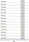
Fig. 4. HPwSTD subspectra of 3-MT (100 μM) and AuNPs (at the overall ligand concentration of 500 μM). Conditions: 500 MHz, 25 °C, [HEPES] = 1 mM, pD = 7.0, H2O/D2O 90:10, 512 scans. Full spectra are reported in Fig. S46.
Fig. 5
Fig. 5. (a) HPwSTD subspectra of 3-MT (25–100 μM) and SO3–AuNP or ProAlaAsp (500 μM, ligand concentration). Conditions: 500 MHz, 25 °C, [HEPES] = 1 mM, pD = 7.0, H2O/D2O 90:10, 512 scans. (b) Signal-to-noise ratio of the 3-MT signal at 6.81 ppm ([3-MT] = 25–100 μM) with SO3–AuNP or ProAlaAsp at 25 μM or 500 μM. Full spectra are reported in Fig. S47.
Fig. 6
Fig. 6. 1H NMR subspectra of 3-MT (50 μM), hippuric acid (50 μM), homovanillic acid (50 μM), vanillic acid (50 μM) and ProAlaAsp (500 μM, ligand concentration). Conditions: 500 MHz, 25 °C, [HEPES] = 1 mM, pD = 7.0, H2O/D2O 90:10). (a) 1H spectrum (128 scans); (b) HPwSTD spectrum (512 scans). Signals at 8.2 ppm belong to amide protons that receive saturation via chemical exchange with H2O. The asterisk denotes a subtraction artifact. The full HPwSTD spectrum is reported in Fig. S48.

References

    1. Sarkar S. Ballester P. Spektor M. Kataev E. A. Angew. Chem., Int. Ed. 2023;62(28):e202214705. doi: 10.1002/anie.202214705. - DOI - PubMed
    1. Li J. Nowak P. Otto S. J. Am. Chem. Soc. 2013;135:9222–9239. doi: 10.1021/ja402586c. - DOI - PubMed
    1. BelBruno J. J. Chem. Rev. 2019;119:94–119. doi: 10.1021/acs.chemrev.8b00171. - DOI - PubMed
    1. Eersels K. Lieberzeit P. Wagner P. ACS Sens. 2016;1:1171–1187. doi: 10.1021/acssensors.6b00572. - DOI
    1. Manhas J. Edelstein H. I. Leonard J. N. Morsut L. Nat. Chem. Biol. 2022;18:244–255. doi: 10.1038/s41589-021-00926-z. - DOI - PMC - PubMed

LinkOut - more resources