. 2025 Feb 12;33(1):101433.
doi: 10.1016/j.omtm.2025.101433. eCollection 2025 Mar 13.

Selective RNAi silencing of Schwann cell Piezo1 alleviates mechanical hypersensitization following peripheral nerve injury

Affiliations

Selective RNAi silencing of Schwann cell Piezo1 alleviates mechanical hypersensitization following peripheral nerve injury

Brandon Itson-Zoske et al. Mol Ther Methods Clin Dev. .

Abstract

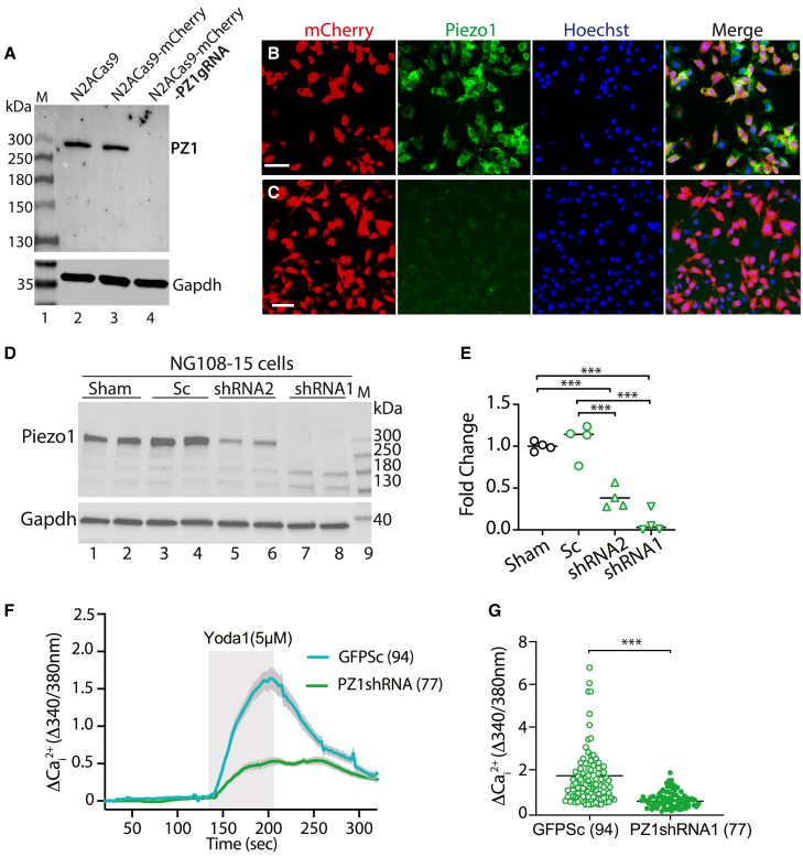
The present study was designed to investigate the role of Schwann cell (SC) Piezo1 in peripheral nociception. We first developed an AAV vector that has primary SC tropism after delivery into the sciatic (or tibial) nerve. This was achieved by packing AAV-GFP transcribed by a CBA promoter using a capsid AAVolig001 to generate AAVolig001-CBA-GFP. Six weeks after intraneural injection of AAVolig001-CBA-GFP in naive rats, GFP expression was detected selectively in both myelinating SCs (mSCs) and non-myelinating SCs (nmSCs). A dual promoter and bidirectional AAV encoding a U6-driven short hairpin RNA against rat Piezo1 (PZ1shRNA) and CBA-transcribed GFP was packed with capsid olig001 (AAVolig001-PZ1shRNA), and AAV was injected into unilateral sciatic (or tibial) nerve immediately after induction of common peroneal nerve injury (CPNI). Results showed that the development of mechanical hypersensitivity in the CPNI rats injected with AAVolig001-PZ1shRNA was mitigated compared to rats subjected to AAVolig001-scramble. Selective in vivo SC transduction and functional block of Piezo1 channel activity of primary cultured SCs was confirmed. These data demonstrate that (1) AAVolig001 has unique and selective primary tropism to SCs via intraneural delivery, and (2) SC Piezo1 contributes to mechanical hypersensitivity following nerve injury.

Keywords: AAVolig001; Piezo1; Schwann cells; intraneural injection; neuropathic pain; peripheral nervous system.


Conflict of interest statement

The authors declare no competing interests.

Figures

None
Graphical abstract
Figure 1
Figure 1
GFP expression after intrasciatic AAVolig001-CBA-GFP (A–D) GFP expression in the nerve fascicles (dashed circles) of cross-sectioned sciatic nerves (A and B). Detection of GFP signals in longitudinal sections of tibial and sural serves (C and D). (E–J) Representative IHC images illustrate selective GFP expression in both mSCs and nmSCs, colabeled with SCs markers MPZ, MBP, GAP43, and P75NTR (E–H). Empty and white arrowheads in (F) and (H) point to mSCs and nmSCs, respectively; no GFP signals in the afferent axons labeled by Tubb3 and NF200 (I and J). Scale bars (μm): (A) and (B), 100; (C), 500; (D)–(F), 100. Antibodies for double labeling are indicated in each montage IHC image. Scales: 50 μm for all. GAP43, growth-associated protein 43; MBP, myelin basic protein; MPZ, myelin protein zero; NF200, neurofilament 200; P75NTR, p75 neurotrophin receptor; and Tubb3, β3-tubulin.
Figure 2
Figure 2
Non-cellular selective transduction after sciatic nerve delivery of AAV6-CBA-GFP (A–F) Representative IHC images illustrate that 5 weeks after sciatic nerve injection of AAV6-CBA-GFP, GFP signals are detected in the afferent axons and SCs of the sciatic nerve by double labeling of GFP with neuronal markers (Tubb3 and NF200) and SC markers (MBP and S100). (F) Representative IHC images show GFP signal detection, colabeled with NF200 in the sensory terminals on the subcutaneous section. (G–I) Neuronal profile of GFP signals in the L3, L4, and L5 DRG ipsilateral to injection. (J–J2) GFP signals are also detected in the spinal cord dorsal horn (DH) neuropil and ventral horn (VH) motor neurons. Scale bars: 50 μm for all.
Figure 3
Figure 3
Cell-based Piezo1 CRISPR knockout and RNAi knockdown (NG108 cells) (A–G) Piezo1 antibody recognizes a clean ∼300-kDa band of canonical Piezo1 protein by immunoblot with comparable band density in control Cas9N2A cells and Cas9N2A cells expressing mCherry, while the Piezo1 band vanishes in the Cas9N2A cells expressing Piezo1-gRNA (A). ICC shows the detection of Piezo1 in Cas9N2A cells expressing mCherry (B), while Piezo1 signals are barely detected in the Cas9N2A cells expressing Piezo1-gRNA (C). Scale bars: 50 μm for (B) and (C). Immunoblots show Piezo1 knockdown after transfection of Piezo1shRNAs into NG108 cells (D and E). Significant reduction of Yoda1-stimulated increase of Cai2+ in NG108 cells expressing Piezo1shRNA1, compared to scramble (Sc) control (F and G).
Figure 4
Figure 4
Mitigation of mechanical hypersensitivity in CPNI rats (A–D) The time courses for the grouped averages to vF, Pin, heat, and cold before (BL) and after sciatic nerve injection of saline or AAVolig001-PZ1shRNA1 in naive rats and after induction CPNI immediately followed by intrasciatic injection of either AAVolig001-PZ1shRNA1 or AAVolig001-Sc. ∗p < 0.05; ∗∗p < 0.01; ∗∗∗p < 0.001 for comparisons to BL within group; #p < 0.05; ##p < 0.01 for comparisons between groups. Repeated-measures parametric two-way ANOVA for vF and heat tests and Tukey’s post hoc test, and Friedman ANOVA for Pin and cold tests and Dunn’s post hoc test. Blue ∗∗∗ is p < 0.001 for main effect comparison. Right sides of (A)–(D) show AUCs calculated using the measures at BL and after vector injection in CPNI rats; ∗p < 0.05; ∗∗p < 0.01; comparisons of AUCs between AAVolig001-PZ1shRNA1 and control vector, unpaired and two-tailed Student’s t test.
Figure 5
Figure 5
Validation of in vivo SCs-Piezo1 knockdown (A–D) Representative IHC montage images of sciatic nerve sections illustrate PZ1 immunopositivity in control vector transduced ring-like SCs (green, arrowheads) and non-transduced axons (red, arrows) (A), while SCs-Piezo1 immunostaining signals (arrowheads) are apparently reduced in AAVolig001-PZ1shRNA1 injected rats (B). PZ1 co-stained with Tubb3 from GFP-Sc (C) and GFP-PZ1shRNA (D). (E–G) Immunoblots show that the Piezo1 protein level in GFP-PZ1shRNA1-expressing primary cultured SCs (E) is significantly reduced as compared to naive and GFP-Sc control (F and G). (H and I) Primary cultured SCs from control animals exhibit an increase in Cai2+ responding to Yoda1 stimulation, while this Yoda1-evoked response is significantly reduced in the SCs expressing PZ1shRNA1 (identified by GFP signals) (H and I). Scale bar (μm): (A-D) 10, (E) 100 and (F) 50.
Figure 6
Figure 6
Tibial injection of AAVolig001-PZ1shRNA1 facilitates skin SCs transduction (A–G) Representative IHC montage images on hindpaw glabrous skin sections after tibial nerve GFP-PZ1shRNA injection. GFP staining (green), colabeled with S100 (red) on the nerve bundles (dashed circles), v denotes vessels (A). GFP staining colabeled with PZ1 in subcutaneous nerve bundles from control vector GFP-Sc (B) and GFP-PZ1shRNA (C); arrowheads and arrows point to axonal PZ1 and SCs-PZ1, respectively. GFP staining colabeled with CK14 (D), S100 (E), and Sox10 (F). (G) Meissner corpuscle colabeled with GFP with Sox10. Scale bars (μm): (A)–(F) 50 and (G) 10. (H and I) The time courses for the grouped averages to vF (H) and Pin (I) at BL and after tibial nerve AAV injection in CPNI rats (n = 8 per group). ∗p < 0.05; ∗∗∗p < 0.001; one-way ANOVA and Tukey post hoc, compared to BL; #p < 0.05; ##p < 0.01; ###p < 0.001, compared between groups; blue ∗∗∗ denotes the comparisons of grouped main effects. Right sides of (H) and (I) are AUCs; ∗∗p < 0.01; ∗∗∗p < 0.001; unpaired and two-tailed Student t tests.

Update of

References

    1. Campana W.M. Schwann cells: activated peripheral glia and their role in neuropathic pain. Brain Behav. Immun. 2007;21:522–527. - PMC - PubMed
    1. Wei Z., Fei Y., Su W., Chen G. Emerging Role of Schwann Cells in Neuropathic Pain: Receptors, Glial Mediators and Myelination. Front. Cell. Neurosci. 2019;13:116. - PMC - PubMed
    1. Gillespie C.S., Sherman D.L., Fleetwood-Walker S.M., Cottrell D.F., Tait S., Garry E.M., Wallace V.C., Ure J., Griffiths I.R., Smith A., Brophy P.J. Peripheral demyelination and neuropathic pain behavior in periaxin-deficient mice. Neuron. 2000;26:523–531. - PubMed
    1. Ding Y.Q., Qi J.G. Sensory root demyelination: Transforming touch into pain. Glia. 2022;70:397–413. - PubMed
    1. De Logu F., Nassini R., Materazzi S., Carvalho Gonçalves M., Nosi D., Rossi Degl'Innocenti D., Marone I.M., Ferreira J., Li Puma S., Benemei S., et al. Schwann cell TRPA1 mediates neuroinflammation that sustains macrophage-dependent neuropathic pain in mice. Nat. Commun. 2017;8:1887. - PMC - PubMed

LinkOut - more resources