. 2025 Feb 27;10(9):9661-9674.
doi: 10.1021/acsomega.4c10956. eCollection 2025 Mar 11.

The Novel Long-Acting Peptide S6-FA Attenuates Liver Fibrosis In Vitro and In Vivo

Affiliations

The Novel Long-Acting Peptide S6-FA Attenuates Liver Fibrosis In Vitro and In Vivo

Mingmin Li et al. ACS Omega. .

Abstract

Liver fibrosis can progress to cirrhosis and hepatocellular carcinoma. Currently, there is no effective drug for liver fibrosis. The peptide 6 (T6) is an endogenous peptide derived from human intrauterine adhesion tissues and has antifibrotic potential. Here, to improve the long-term efficacy and activity of T6, we conducted the rational modified of T6 through studying structure-activity, and synthesized a series of analogues. Among them, S6 and S6-FA exhibited optimal antihepatic fibrosis activity, and S6-FA had a stronger long-acting effect than T6 and S6. The two analogues inhibited the expression of α-SMA and Collagen 1 in TGF-β-induced LX2 cells model and CCl4-induced mouse model of liver fibrosis. Besides, we discovered that S6 and S6-FA remarkably reduced the AST and ALT serum levels. Mechanistic studies have demonstrated that analogues inhibited liver fibrosis through inhibiting Erk, Smad and P65 pathways. This study provided that the novel peptide S6 and S6-FA is potential candidate compounds for treating liver fibrosis.


Conflict of interest statement

The authors declare no competing financial interest.

Figures

Figure 1
Figure 1
Characterization of the peptides. (A) Western blot analysis of FN and COL1α1 expression level in TGF-β-induced LX2 cells treated with different concentrations of peptide T6 (n = 3). (B) Alanine scanning mutagenesis of T6 and corresponding peptides against inhibited the expression of FN and COL1α1, as measured by Western blot assay. (C) Western blot analysis of FN and COL1α1 expression in a TGF-β-induced cell model treated with 10 μΜ analogues (n = 3). Data are represented as mean ± SEM *P < 0.05, **P < 0.01, ***P < 0.001 and ****P < 0.0001. All compounds are >95% pure by HPLC analysis.
Figure 2
Figure 2
Analysis of the properties of truncated peptides and the conformation of T6-S. (A) Names and sequences of truncated peptides. (B-E) Western blot analysis of FN and COL1α1 expression level in TGF-β-induced LX2 cells treated with different concentrations of short peptide (n = 3). (F) Optimal structures of T6-S in water generated from MD simulations. (G) Peptide T6-S targeting mitochondrial membrane for fluorescence confocal microscopy. Data are represented as mean ± SEM **P < 0.01, ***P < 0.001 and ****P < 0.0001. All compounds are >95% pure by HPLC analysis.
Figure 3
Figure 3
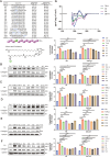
Characterization of the peptides. (A) Inhibition of FN and COL1α1 expression in LX2 cells as determined by Western blot analysis (n = 3). (B-E) The modifications of maternal peptide T6-S and the antifibrosis effect of S1–S19 peptides in vitro (n = 3). (F) Intensive screening of modified peptides for enhanced therapeutic effects, with detection of FN and COL1α1 expression (n = 3). (G) CD spectra of T6-S, S5, S6 and S6-FA in 50% trifluoroethanol (TFE) aqueous solution. Data are represented as mean ± SEM *P < 0.05, **P < 0.01, ***P < 0.001 and ****P < 0.0001. All compounds are >95% pure by HPLC analysis.
Scheme 1
Scheme 1. Synthesis of Peptides T6-S
Reagents and conditions: (a) N,N-dimethylformamide (DMF) + dichloromethane (DCM), 25 °C, 0.5 h. (b) Fmoc-Ser(t-Bu)–OH, PYBOP, N,N′-diisopropylcarbodiimide (DIC), N,N-diisopropylamine (DIEA), DMF, 25 °C, 4 h. (c) 20% piperidine/DMF, 25 °C, 6 + 8 min. (d) Fmoc-Xaa–OH, 1-hydroxybenzotriazole (HOBt), N,N′-diisopropylcarbodiimide (DIC), DMF, 25 °C, 2 h. (e) Trifluoroacetic acid (TFA)/triisopropylsilane (TIS)/H2O = 95:2.5:2.5 (v/v/v), 0 → 25 °C, 2 h.
Figure 4
Figure 4
Validate the fibrosis pathway and its effects on phenotypic proteins in vitro. (A) Western blot analysis of p-ERK, p-Smad3, p-P65, α-SMA and COL1α1 in LX2 cells. Cells were treated with TGF-β or candidate peptides (10 μM) for 24 h. GAPDH was used as a loading control. (B) Densitometric analysis of the ERK, Smad3, and P65 phosphorylation in (A) (n = 3). (C) Densitometric analysis of the α-SMA, and COL1α1 in (A) (n = 3). Data are represented as mean ± SEM *P < 0.05, **P < 0.01, ***P < 0.001 and ****P < 0.0001.
Figure 5
Figure 5
In vivo study of candidate peptides in a CCl4-induced liver fibrosis model. (A) Schematic illustration of the CCl4-indcued liver fibrosis model and the in vivo treatment procedure. (B) Histological images of hepatic tissues by H&E, Sirius red staining and immunohistochemical (IHC) staining for α-SMA, COL1α1(magnification: 20 ×; n = 5). (C) Monitoring of changes in mouse body weight (n = 5–7). (D) Positive staining area analyses of the histological images shown in (B). (E) Analyses of hydroxyproline in liver tissue (n = 5). (F, J) Analyses of ALT and AST in serum (n = 5). (H) Analysis of Positive Staining Areas in Histological Images by Immunohistochemistry shown in (B). (I, J) The serum stability of maternal peptide T6-S and S6-FA. Data are represented as mean ± SEM **P < 0.01, ***P < 0.001 and ****P < 0.0001.
Figure 6
Figure 6
Validate the fibrosis pathway and its impact on phenotypic proteins, and improve oxidative stress in vivo. (A) Western blot analysis of p-ERK, p-Smad3, p-P65, α-SMA and COL1α1 in Histological tissue of the liver. (B) Densitometric analysis of the ERK, Smad3, and P65 phosphorylation in (A) (n = 5). (C) Densitometric analysis of the α-SMA, and COL1α1 in (A) (n = 5). (DE) The level of SOD and GSH based on enzyme activity assay (n = 5). Data are represented as mean ± SEM **P < 0.01, ***P < 0.001 and ****P < 0.0001.

References

    1. Horn P.; Tacke F. Metabolic reprogramming in liver fibrosis. Cell Metab. 2024, 36 (7), 1439–1455. 10.1016/j.cmet.2024.05.003. - DOI - PubMed
    1. Yan Z.; Cheng X.; Wang T.; Hong X.; Shao G.; Fu C. Therapeutic potential for targeting Annexin A1 in fibrotic diseases. Genes Dis. 2022, 9 (6), 1493–1505. 10.1016/j.gendis.2022.05.038. - DOI - PMC - PubMed
    1. Parola M.; Pinzani M. Liver fibrosis: Pathophysiology, pathogenetic targets and clinical issues. Mol. Aspects Med. 2019, 65, 37–55. 10.1016/j.mam.2018.09.002. - DOI - PubMed
    1. Pellicoro A.; Ramachandran P.; Iredale J. P.; Fallowfield J. A. Liver fibrosis and repair: immune regulation of wound healing in a solid organ. Nat. Rev. Immunol. 2014, 14 (3), 181–194. 10.1038/nri3623. - DOI - PubMed
    1. Xu H.; Zhao Q.; Song N.; Yan Z.; Lin R.; Wu S.; Jiang L.; Hong S.; Xie J.; Zhou H.; Wang R.; Jiang X. AdipoR1/AdipoR2 dual agonist recovers nonalcoholic steatohepatitis and related fibrosis via endoplasmic reticulum-mitochondria axis. Nat. Commun. 2020, 11 (1), 580710.1038/s41467-020-19668-y. - DOI - PMC - PubMed

LinkOut - more resources