. 2025 Feb 21.
doi: 10.1039/d4md01016g. Online ahead of print.

Design, synthesis and biological evaluation of 2 H-[1,4]oxazino-[2,3- f]quinazolin derivatives as potential EGFR inhibitors for non-small cell lung cancer

Affiliations

Design, synthesis and biological evaluation of 2 H-[1,4]oxazino-[2,3- f]quinazolin derivatives as potential EGFR inhibitors for non-small cell lung cancer

Linchang Huang et al. RSC Med Chem. .

Abstract

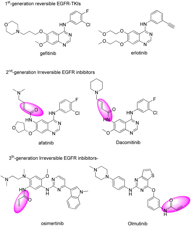
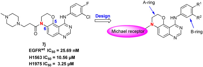
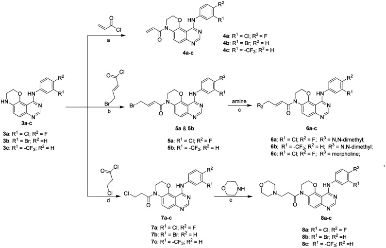
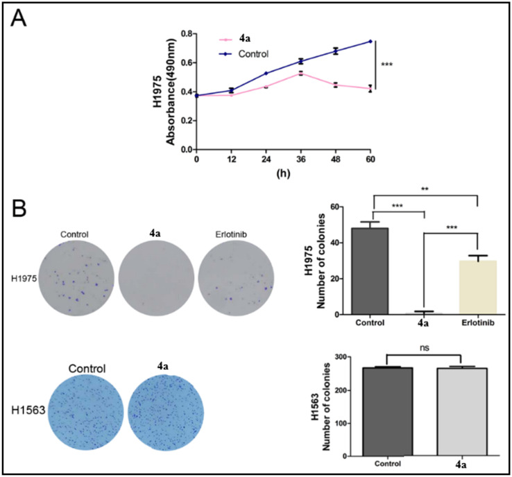
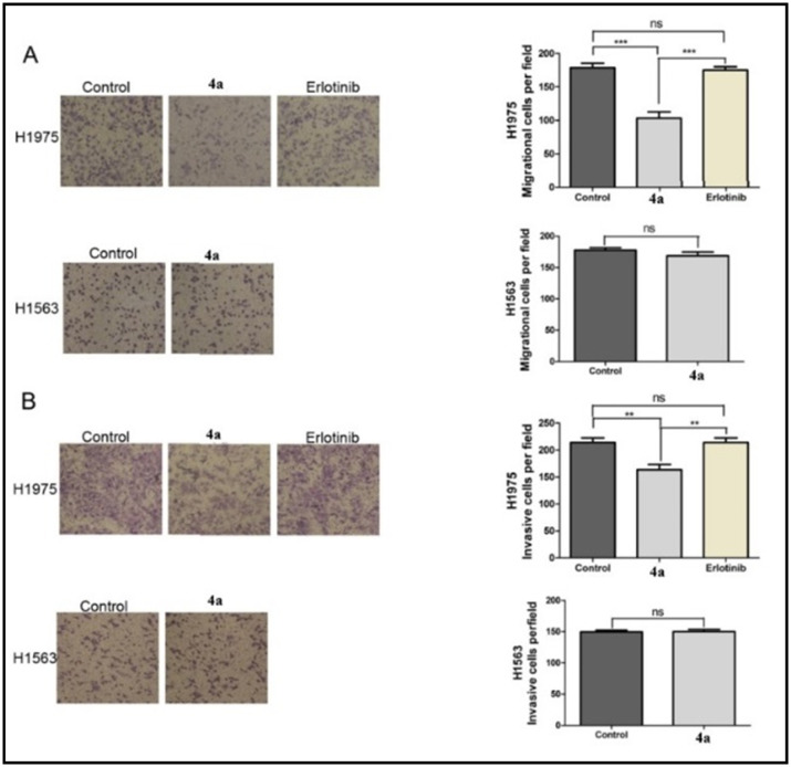
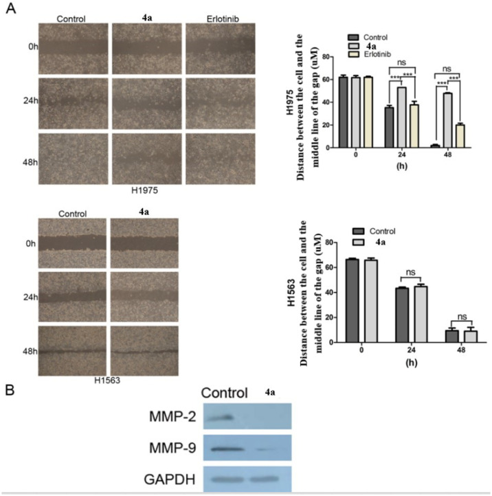
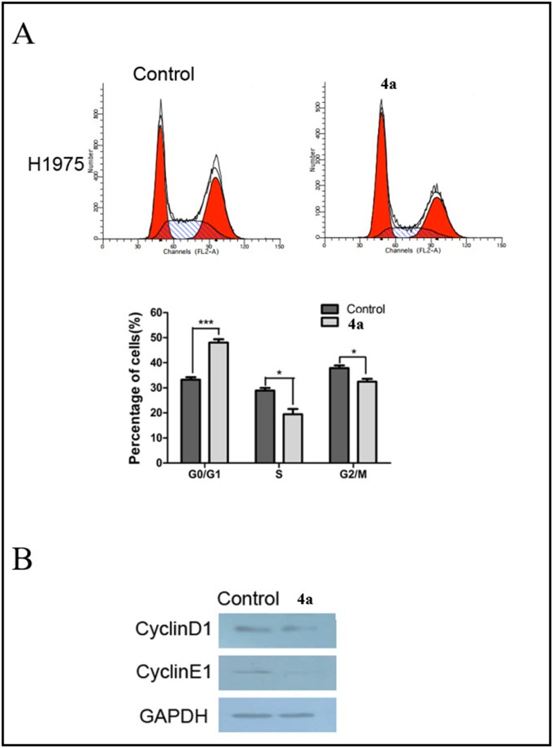
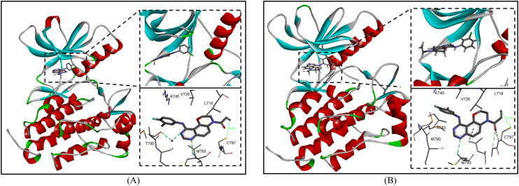
Epidermal growth factor receptor tyrosine kinase inhibitors (EGFR-TKIs) have emerged as the first-line treatment for patients with EGFR-mutant non-small cell lung cancer (NSCLC). A series of 2H-[1,4]oxazino[2,3-f]quinazolin derivatives were synthesized and evaluated as irreversible EGFR-TKIs for the treatment of NSCLC. Most of the synthesized compounds demonstrated strong inhibitory activity against the EGFR kinase and the tested cancer cells. Notably, compound 4a exhibited considerable inhibitory effects against the EGFR kinase and the EGFRL858R/T790M mutant NCI-H1975 cancer cells. Compound 4a was found to suppress cell proliferation, colony formation, cell invasion, and migration, while also inducing G0/G1 phase arrest of the cell cycle in NCI-H1975 cells. Compound 4a was docked into the active pocket of the EGFR mutant to ascertain the probable binding conformation. Overall, compound 4a was identified as a promising irreversible EGFR-TKI for the treatment of NSCLC.


Conflict of interest statement

The authors declare no conflicts of interest.

Figures

Fig. 1
Fig. 1. Development of three generations of small-molecule EGFR-TKIs (the second-generation and third-generation EGFR inhibitors contained an electrophilic Michael acceptor).
Fig. 2
Fig. 2. Design strategy and modification of novel EGFR inhibitors.
Scheme 1
Scheme 1. Synthesis of target compounds. Reagents and reaction conditions: (a) THF, 50 °C; (b) DMF/DCM, NEt3, 0 °C; (c) K2CO3, DMF, rt; (d) THF, rt; (e) KI, ethanol, reflux.
Fig. 3
Fig. 3. (A) The impact of compound 4a on H1975 cell viability was assessed using the MTT assay over a 60-hour period. This experiment was conducted three times, showing statistically significant results (***P < 0.001). (B) Colony formation assays indicate that the number of clones decreases after treatment with compound 4a and erlotinib (0.5 μM) in H1975 cells. However, treatment with compound 4a did not alter clone formation in H1563 cells. The average number of tumor clones is presented in the bar diagram. Results are statistically significant as follows: ***P < 0.001, **P < 0.01, ns (not significant).
Fig. 4
Fig. 4. Compound 4a suppresses lung cancer cell invasion and migration (Transwell). (A) Cell migration in H1975 and H1563 cells was assessed before and after treatment with compound 4a (0.5 μM), as well as in H1975 cells before and after treatment with erlotinib. The migratory cell numbers are presented in a bar diagram. Each experiment was repeated three times. ***P < 0.001, ns, ns. (B) Cell invasion in H1975 and H1563 cells was evaluated before and after treatment with compound 4a (0.5 μM), as well as in H1975 cells before and after treatment with erlotinib. The number of cells invading the lower chamber is shown in a bar diagram. Each experiment was repeated three times. **P < 0.01, ns, ns.
Fig. 5
Fig. 5. Wound healing and western blot assay. (A) Wound healing assay results in H1975 and H1563 cells before and after treatment with compound 4a (0.5 μM), as well as in H1975 cells before and after treatment with erlotinib. The migratory cell number is presented by bar diagram. Each experiment was repeated three times. ***P < 0.001, ns, ns. (B) The expression levels of MMP2 and MMP9 were detected by western blot.
Fig. 6
Fig. 6. The effect of compound 4a on H1975 cell cycle. (A) Cell phase distribution in H1975 cells before and after treatment with compound 4a (0.5 μM). The ratio is presented by bar diagram. Each experiment was repeated three times. ***P < 0.001. (B) The expression of cyclinD1 and cyclinE1 was detected by western blot in H1975 cells before and after treatment with compound 4a. GAPDH: internal control.
Fig. 7
Fig. 7. (A) Compound 4a binding into the active site of wild-type EGFR kinase (PDB code: 1M17; binding energy: −7.35 kcal mol−1); (B) 2D model of compound 4a binding into the active site of EGFRT790 kinase (PDB code: 4G5P; binding energy: −8.27 kcal mol−1).

Similar articles

References

    1. Sung H. Ferlay J. Siegel R. L. Laversanne M. Soerjomataram I. Jemal A. Bray F. Ca-Cancer J. Clin. 2021;71:209. - PubMed
    1. Das D. Xie L. Hong J. RSC Med. Chem. 2024;15:3371. - PMC - PubMed
    1. Herbst R. S. Morgensztern D. Boshoff C. Nature. 2018;553:446. - PubMed
    1. Das D. Xie L. Wang J. Shi J. Hong J. Bioorg. Chem. 2020;99:103790. - PubMed
    1. Warnecke B. Nagasaka M. Adv. Oncol. 2024;4:63.