. 2025 Mar 13.
doi: 10.1039/d4md00698d. Online ahead of print.

Development, biological evaluation, and molecular modelling of novel isocytosine and guanidine derivatives as BACE1 inhibitors using a fragment growing strategy

Affiliations

Development, biological evaluation, and molecular modelling of novel isocytosine and guanidine derivatives as BACE1 inhibitors using a fragment growing strategy

Asmaa M Atta et al. RSC Med Chem. .

Abstract

Alzheimer's disease (AD) is a neurodegenerative condition characterized by significant synaptic loss and neuronal death in brain regions critical for cognitive functions. The disease is characterized by the formation of amyloid plaques, which are extracellular constructs consisting mainly of aggregated Aβ42. The latter is a peptide formed by the proteolytic cleavage of β-amyloid precursor protein (APP) by two enzymes, β- and γ-secretase. Therefore, inhibition of the aspartic protease β-secretase (BACE1) is considered a promising therapeutic approach for the treatment and prevention of Alzheimer's disease. Unfortunately, a limited number of β-secretase inhibitors have reached human trials and eventually failed due to inconclusive therapeutic and/or safety profiles. In this study, we developed drug-like molecules with a β-secretase inhibitory activity using a fragment growing strategy on isocytosine and acyl guanidine warheads. Our approach is based on optimizing the hydrophobic part of the molecules to obtain a conformationally restrained scaffold complementary to the hydrophobic pockets within the enzyme active site. We developed 32 compounds with promising in vitro inhibitory activity against BACE1 down to sub-micromolar IC50. Docking simulation studies were performed to understand the mode of binding of the prepared compounds. We demonstrated that compounds with superior activities, such as 16b and 16g, are able to provide the best balance between the steric shape and position of the polar substituent for achieving preferential anchoring into the S1, S3, S1', and S2' sub-pockets. Further, in vivo characterization of selected drug-like candidates of the benzimidazole series AMK-IV, namely 16a and 16k, demonstrated their ability to reduce oxidation stress and their safety within brain and liver tissues.


Conflict of interest statement

The authors declare no competing interest.

Figures

Fig. 1
Fig. 1. Pathophysiology of AD highlighting the core disease pathologies. Formation of β-amyloid plaques and tau hyperphosphorylated neurofibrillary tangles are the disease's main microscopical examination findings. The associated hypothesis of neuroinflammatory signaling and mitochondria-driven oxidative stress is associated and/or correlated with β-amyloid and hyperphosphorylated tau formation.
Fig. 2
Fig. 2. Chemical structures of currently known BACE1 inhibitors with clinical trial records.
Fig. 3
Fig. 3. Rational design of our BACE-1 inhibitors. (A) Structure of human BACE-1 (PDB: 2QU3) represented in gray cartoon highlighting the main ligand's hydrophobic binding sub-pockets (S1, S3, S1′, and S2′) as well as key protein secondary structures (flap, 10S loop, loops C, D, F). (B) Schematic representation for the pharmacophoric features of inhibitors targeting the BACE-1 catalytic binding site. (C) Structure- and ligand-based drug design for our synthesized compounds based on warhead isosteric replacement, atom interconnection for ring addition, central ring hopping, linker expansion, and projections into hydrophobic sub-pockets.
Scheme 1
Scheme 1. Synthetic route for preparation of AMK-I and AMK-II analogues. Reagents and conditions: (a) CDI, MgCl2, potassium malonate, Et3N, ACN, rt, overnight; (b) guanidine, sodium ethoxide, ETOH, 80 °C, overnight; (c) Ar–B(OH)2, Pd(PPh3)4, 1 M K2CO3, toluene, MeOH, 100 °C, 48 h.
Scheme 2
Scheme 2. Synthetic route for preparation of AMK-III analogues. Reagents and conditions: (a) POCl3, DMF, rt, 2 h; (b) Ar–B(OH)2, Pd(PPh3)4, MeOH, toluene, 100 °C, 48 h; (c) KMnO4, acetone, 5 h; (d) CDI, guanidine, NaH, 5 h.
Scheme 3
Scheme 3. Synthetic route for preparation of AMK-IV analogues. Reagents and conditions: (a) RBr or ArBr, K2CO3, DMF, rt, 24 h; (b) synthesized 13a–i or the commercially available 13j and k, phenylenediamine, NH4Cl, EtOH, 80 °C, 4 h. (c) Ethyl bromo acetate, NaH, DMF, rt, 24 h; (d) guanidine, NaH, DMF, 70 °C, 17 h.
Fig. 4
Fig. 4. Insights from the structural–activity relationship of the synthesized compounds as per their inhibition activity against the hBACE-1 enzyme.
Fig. 5
Fig. 5. Proposed binding modes of selected compounds into the crystal structure of the BACE1 enzyme (PDB ID: 2QU3). The compounds depicted are 4a (A), 4′g (B), 9a (cyan) and 9c (purple) overlaid (C), and 16b (cyan) and 16g (purple) overlaid (D). Ligands are shown as sticks, while the enzyme active site is shown as a surface. Figures were prepared using the Pymol molecular graphics software.
Fig. 6
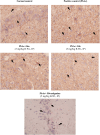
Fig. 6. Photomicrographs of brain tissue of different experimental groups stained by β-amyloid using immunohistochemical staining (magnification of 40×). Weak expression in neuronal tissue (normal control), strong expression in neuronal tissue (PbAc control), moderate expression in neuronal tissue (16a-treated), weak expression in neuronal tissue (16k-treated), and significant reduction in the expression of β-amyloid (black arrows in the rivastigmine-treated group).
Fig. 7
Fig. 7. Assessment of oxidant and antioxidant levels in different treated groups; (A) oxidant: malondialdehyde (MDA) and nitric oxide (NO) and (B) antioxidant: reduced glutathione (GSH), catalase (CAT) and superoxide dismutase (SOD) activities in normal, untreated, and treated PbAc-induced (neurotoxicity) rats. * Values are significantly different (P ≤ 0.05) between positive control and normal groups, while # (P ≤ 0.05) values are significantly different between untreated and treated-PbAc-induced rats (neurotoxicity) using the unpaired test in GraphPad Prism.
Fig. 8
Fig. 8. Left panel: Histopathological examinations of brain tissue of rats in different experimental groups stained with H&E (magnification of 40×). Normal neurons (black arrows) within background of glial tissue. Positive control (PbAc); there is a reduction in the number of neurons (black arrows), with many red neurons (black arrowheads). There is edema (blue arrows). There are scattered chronic inflammatory cell infiltrates (red arrowheads). There is extensive gliosis (red arrows). PbAc + 16a; there is a partial restoration of neuronal tissue (black arrows), reduction in red neurons (black arrowheads), edema (blue arrows) and chronic inflammatory cells (red arrowheads). There is still gliosis (red arrows). PbAc + 16k and rivastigmine-treated; there is almost restoration of neuronal tissue (black arrows), with scattered few red neurons (black arrowheads). There is no edema or chronic inflammatory cell infiltrate or gliosis (H&E, 40×). Right panel: Bar representation for the quantification of the number of neurons in different treated groups. *P ≤ 0.05: values are significantly different between untreated and treated PbAc-induced rats (neurotoxicity) using the unpaired test in GraphPad Prism. Normal control: grade (0), Pb-Ac control: grade (3 = 80%), compound 16a-treated Pb-Ac rats: grade (2 = 35%), compound 16k-treated Pb-Ac rats: grade (1 = 5%) and rivastigmine-treated Pb-Ac rats: grade (1 = 5%). The grades were scaled regarding red neurons, edema, chronic inflammatory cells, and gliosis. The severity of microscopic lesions observed was graded based on the degree and extent of tissue damage using a four-point scale modified from Jokinen et al. (2011).
Fig. 9
Fig. 9. Histopathological examination of liver tissues in treated groups (H&E, 40×). Normal control with uniform liver tissue, regular plates of hepatocytes, and no evidence of injury (black arrow heads). Positive control with severe hydropic degeneration in hepatocytes (black arrow heads). There are foci of lytic necrosis (red arrows) in positive control rats. PbAc rats treated with compounds 16a and 16k, with mild hydropic degeneration in hepatocytes (black arrow heads), with one focus of lytic necrosis (red arrows) in the 16a and 16k-treated rats. In the rivastigmine–PbAc treated rats, hepatocytes show evidence of hydropic degeneration (black arrowheads). There are foci of lytic necrosis (red arrows). There is mild portal tract inflammation (black arrow).
Fig. 10
Fig. 10. Experimental design for the lead acetate induced neurotoxicity in male Wistar rats.

References

    1. Knopman D. S. Jack, Jr. C. R. Wiste H. J. Weigand S. D. Vemuri P. Lowe V. J. Kantarci K. Gunter J. L. Senjem M. L. Mielke M. M. Machulda M. M. Roberts R. O. Boeve B. F. Jones D. T. Petersen R. C. Neurology. 2016;87:691–698. - PMC - PubMed
    1. Wilson R. S. Hoganson G. M. Rajan K. B. Barnes L. L. Mendes de Leon C. F. Evans D. A. Neurology. 2010;75:21–26. - PMC - PubMed
    1. Alzheimer's disease facts and figures, Alzheimer's Dementia, 2024, vol. 20(5), pp. 3708–3821 - PMC - PubMed
    1. Skaria A. P. Am. J. Manag. Care. 2022;28(10 Suppl):S188–S196. - PubMed
    1. Deb A. Thornton J. D. Sambamoorthi U. Innes K. Expert Rev. Pharmacoeconomics Outcomes Res. 2017;17:189–202. - PMC - PubMed

LinkOut - more resources