. 2025 Mar 14;31(10):101014.
doi: 10.3748/wjg.v31.i10.101014.

Lactococcus garvieae aggravates cholestatic liver disease by increasing intestinal permeability and enhancing bile acid reabsorption

Affiliations

Lactococcus garvieae aggravates cholestatic liver disease by increasing intestinal permeability and enhancing bile acid reabsorption

Man Liu et al. World J Gastroenterol. .

Abstract

Background: Although an association between gut microbiota and cholestatic liver disease (CLD) has been reported, the precise functional roles of these microbes in CLD pathogenesis remain largely unknown.

Aim: To explore the function of gut microbes in CLD pathogenesis and the effects of gut microbiota on intestinal barrier and bile acid (BA) metabolism in CLD.

Methods: Male C57BL/6J mice were fed a 0.05% 3,5-diethoxycarbonyl-1,4-dihydrocollidine diet for 2 weeks to induce CLD. The sterile liver tissues of mice were then meticulously harvested, and bacteria in homogenates were identified through culture methods. Furthermore, 16S ribosomal DNA sequencing was employed to analyze sterile liver samples collected from eight patients with primary biliary cholangitis (PBC) and three control individuals with hepatic cysts. The functional roles of the identified bacteria in CLD pathogenesis were assessed through microbiota transfer experiments, involving the evaluation of changes in intestinal permeability and BA dynamics.

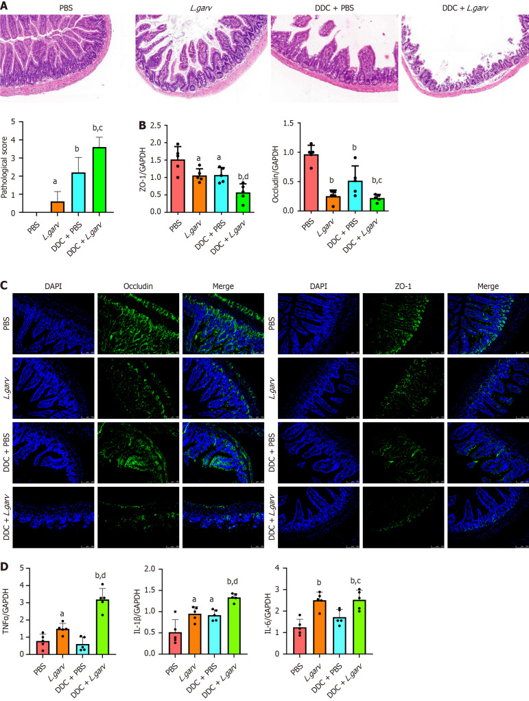
Results: Ligilactobacillus murinus (L. murinus) and Lactococcus garvieae (L. garvieae) were isolated from the bacterial culture of livers from CLD mice. L. murinus was prevalently detected in PBC patients and controls, whereas L. garvieae was detected only in patients with PBC but not in controls. Mice inoculated with L. garvieae exhibited increased susceptibility to experimental CLD, with both in vitro and in vivo indicating that L. garvieae disrupted the intestinal barrier function by down-regulating the expression of occludin and zonula occludens-1. Moreover, L. garvieae administration significantly upregulated the expression of the apical sodium-dependent BA transporter in the terminal ileum and increased serum BA levels.

Conclusion: L. garvieae contributes to excessive BA-induced hepatobiliary injury and liver fibrosis by increasing intestinal permeability and enhancing BA reabsorption.

Keywords: Lactococcus garvieae; Bile acid; Cholestasis; Intestinal permeability; Microbiota.


Conflict of interest statement

Conflict-of-interest statement: All the authors report no relevant conflicts of interest for this article.

Figures

Figure 1
Figure 1
Lactococcus garvieae enrichment in the liver during cholestatic liver disease. A: Bacterial cultures from mouse liver homogenate smears after 2 weeks of feeding chow and 3,5-diethoxycarbonyl-1,4-dihydrocollidine diets; B: The 16S V3-V4 sequencing results were subjected to BLAST to identify specific bacterial species; C: The heat map displays the relative abundance of various bacterial phyla in liver tissues of patients with primary biliary cholangitis (PBC) and controls; D: At the genus level, the relative abundance of Ligilactobacillus and Lactococcus in liver tissues of patients with PBC and controls; E: At the species level, the relative abundance of Ligilactobacillus murinus and Lactococcus garvieae in liver tissues of patients with PBC and controls. PBC: Primary biliary cholangitis; CK: Controls.
Figure 2
Figure 2
Lactococcus garvieae colonization aggravates 3,5-diethoxycarbonyl-1,4-dihydrocollidine-induced cholestatic cholangitis. A: Experimental flow chart; B: Representative images of liver sections stained with hematoxylin and eosin (original magnification, 100 × in the left panels and 200 × in the right panels; scale bars, 100 µm); C: Changes in body weight following the diet switch and Lactococcus garvieae treatment; D: Serum alkaline phosphatase, gamma-glutamyl transferase and alanine aminotransferase levels from each group are presented. All values are expressed as the mean ± SD; aP < 0.05 vs PBS group; bP < 0.01 vs PBS group; cP < 0.05 vs DDC + PBS group; DDC: 3,5-diethoxycarbonyl-1,4-dihydrocollidine; PBS: Phosphate-buffered saline; L. garv: Lactococcus garvieae; ALT: Alanine aminotransferase; ALP: Serum alkaline phosphatase; GGT: Gamma-glutamyl transferase.
Figure 3
Figure 3
Lactococcus garvieae aggravates the impairment of intestinal barrier integrity. A: Representative images of ileum sections stained with hematoxylin and eosin (original magnification 200 ×; scale bars, 50 µm); B: Occludin and zonula occludens-1 (ZO-1) mRNA levels in the ileum of each group; C: Occludin and ZO-1 protein expressions detected using immunofluorescence; D: Tumor necrosis factor alpha (TNFα), interleukin (IL)-6 and IL-1β mRNA levels in the ileum of each group. All values are shown as mean ± SD. aP < 0.05 vs phosphate-buffered saline (PBS) group; bP < 0.01 vs PBS group; cP < 0.05 vs 3,5-diethoxycarbonyl-1,4-dihydrocollidine (DDC) + PBS group; dP < 0.01 vs DDC + PBS group; DDC: 3,5-diethoxycarbonyl-1,4-dihydrocollidine; PBS: Phosphate-buffered saline; L. garv: Lactococcus garvieae; TNFα: Tumor necrosis factor alpha; IL: Interleukin; GAPDH: Glyceraldehyde 3-phosphate dehydrogenase; ZO-1: Zonula occludens-1.
Figure 4
Figure 4
Lactococcus garvieae aggravates hepatic inflammation and fibrosis. A: Representative images of liver sections stained with sirius red (original magnification 200 ×; scale bars, 100 µm); B and C: Α-SMA, COL1A1, COL1A2, COL3A1, COL4A2, TGFβ, TNFα, CCL2, CCL5, CCL20, and P16 mRNA levels in the livers of each group. All values are expressed as the mean ± SD; aP < 0.05 vs phosphate-buffered saline (PBS) group; bP < 0.01 vs PBS group; cP < 0.05 vs 3,5-diethoxycarbonyl-1,4-dihydrocollidine (DDC) + PBS group; dP < 0.01 vs DDC + PBS group; DDC: 3,5-diethoxycarbonyl-1,4-dihydrocollidine; PBS: Phosphate-buffered saline; L. garv: Lactococcus garvieae; TNFα: Tumor necrosis factor alpha; GAPDH: Glyceraldehyde 3-phosphate dehydrogenase.
Figure 5
Figure 5
Ligilactobacillus murinus induces barrier injury and inflammatory activation in vitro. A: Occludin and zonula occludens-1 (ZO-1) mRNA levels in human intestinal epithelial Caco-2 cells; B: Protein ZO-1 and occludin protein levels in Caco-2 cells analyzed using western blotting. Glyceraldehyde 3-phosphate dehydrogenase served as the loading control; C and D: Tumor necrosis factor alpha (TNFα), CCL2, CCL5 and interleukin-1β mRNA levels in RAW264.7 cells (C) and TNFα, CCL2 and CCL5 mRNA levels in human intrahepatic bile duct epithelial cells (D) exposed at different concentrations of Lactococcus garvieae culture supernatants (1:100, 1:50, 1:10). All values are expressed as the mean ± SD; aP < 0.05 vs vehicle group; bP < 0.01 vs vehicle group; ZO-1: Zonula occludens-1; TNFα: Tumor necrosis factor alpha; IL: Interleukin; GAPDH: Glyceraldehyde 3-phosphate dehydrogenase.
Figure 6
Figure 6
Bile acids transportation is altered by Lactococcus garvieae. A: Serum TBA values from each group are presented; B: Apical sodium-dependent bile acid transporter mRNA levels in the intestine of each group; C and D: OATP, NTCP, BSEP, MDR1, cytochrome P450 family 7 subfamily A member 1, CYP8B1 and cytochrome P450 family 27 subfamily A member 1 mRNA levels in the livers of each group; E: Liver TC values from each group are presented. All values are expressed as the mean ± SD; aP < 0.05 vs phosphate-buffered saline (PBS) group; bP < 0.01 vs PBS group; cP < 0.05 vs 3,5-diethoxycarbonyl-1,4-dihydrocollidine (DDC) + PBS group; dP < 0.01 vs DDC + PBS group. PBS: Phosphate-buffered saline; L. garv: Lactococcus garvieae; DDC: 3,5-diethoxycarbonyl-1,4-dihydrocollidine; ASBT: Apical sodium-dependent bile acid transporter; GAPDH: Glyceraldehyde 3-phosphate dehydrogenase; CYP27A1: Cytochrome P450 family 27 subfamily A member 1; CYP7A1: Cytochrome P450 family 7 subfamily A member 1.

References

    1. Jansen PL, Ghallab A, Vartak N, Reif R, Schaap FG, Hampe J, Hengstler JG. The ascending pathophysiology of cholestatic liver disease. Hepatology. 2017;65:722–738. - PubMed
    1. Schaap FG, Trauner M, Jansen PL. Bile acid receptors as targets for drug development. Nat Rev Gastroenterol Hepatol. 2014;11:55–67. - PubMed
    1. Griffiths L, Dyson JK, Jones DE. The new epidemiology of primary biliary cirrhosis. Semin Liver Dis. 2014;34:318–328. - PubMed
    1. Tang R, Wei Y, Li Y, Chen W, Chen H, Wang Q, Yang F, Miao Q, Xiao X, Zhang H, Lian M, Jiang X, Zhang J, Cao Q, Fan Z, Wu M, Qiu D, Fang JY, Ansari A, Gershwin ME, Ma X. Gut microbial profile is altered in primary biliary cholangitis and partially restored after UDCA therapy. Gut. 2018;67:534–541. - PubMed
    1. Chen LP, Zhao H, Lyu B, Cheng JL. [Environmental factors and primary biliary cirrhosis] Zhonghua Gan Zang Bing Za Zhi. 2016;24:541–544. - PubMed

MeSH terms

Substances

LinkOut - more resources