. 2025 Feb 26;22(7):1504-1515.
doi: 10.7150/ijms.102967. eCollection 2025.

Interleukin 26 attenuates osteoblast differentiation in osteoarthritis patients by activating COX2 and NF-κB pathways

Affiliations

Interleukin 26 attenuates osteoblast differentiation in osteoarthritis patients by activating COX2 and NF-κB pathways

Yi-Hsuan Lin et al. Int J Med Sci. .

Abstract

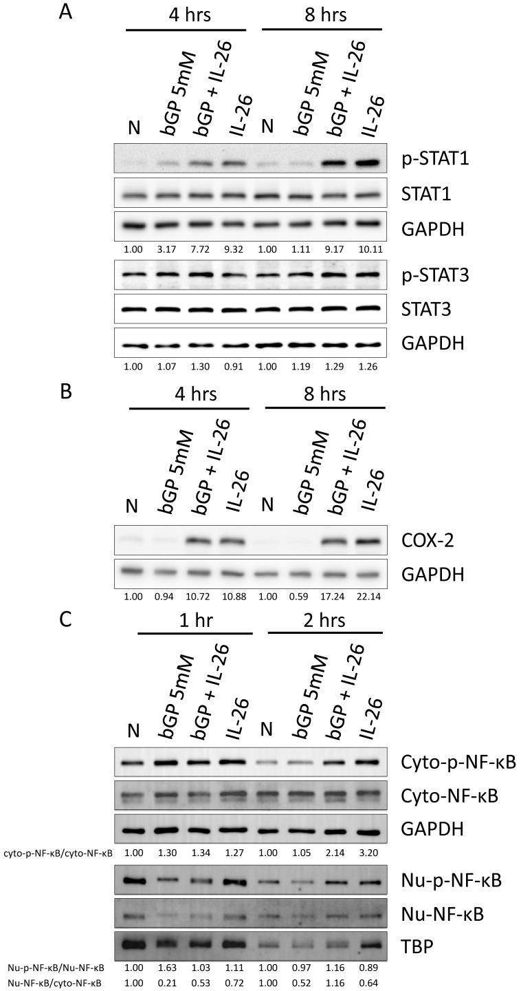
Aims: Osteoarthritis (OA) represents the prevailing form of degenerative joint pathology. Recent investigations have revealed a heightened expression of interleukin 26 (IL-26) in various inflammatory arthritic conditions, including OA. However, the specific impacts and functions of IL-26 on osteoblasts (OBs) within the context of OA remain inadequately elucidated. This study aims to clarify the effects and underlying mechanisms of IL-26 by examining its influence on osteoblasts isolated from OA patients and a murine osteoblast cell line. Methods: Human primary osteoblasts and mouse pre-osteoblast cells were subjected to treatment with β-glycerophosphate or concurrent treatment with IL-26 to observe the effects on osteoblast differentiation. The differentiation of osteoblasts was assessed through the expression of relevant genes using reverse transcription-polymerase chain reaction (RT-PCR). Key molecular mechanisms of downstream signaling pathways were examined through immunoblotting assays. Results: Our results reveal that IL-26 mitigates osteoblast differentiation and reduces the expression of the marker alkaline phosphatase. Furthermore, the NF-κB downstream OB proliferated marker iNOS and inhibition OB differentiated marker LCN2 messenger RNA are up-regulated in IL-26 treated group. Also, phosphorylation and nuclear translocation of NF-κB p65 occur following IL-26 stimulation. Additionally, IL-26 enhances the downstream transcription factor cyclooxygenase-2 (COX2), a major player associated with iNOS. STAT1, the canonical receptor signaling pathway of IL-26 is activated. Conclusion: In summary, our findings substantiate the role of IL-26 in osteoarthritis and identify it as a potential therapeutic target for intervention in osteoarthritic pathology.

Keywords: interleukin 26; osteoarthritis; osteoblast differentiation.


Conflict of interest statement

Competing Interests: The authors have declared that no competing interest exists.

Figures

Figure 1
Figure 1
IL-26 levels in osteoarthritis patients and impact on osteoblast cell viability. (A) Bone sample specimens were obtained from patients with osteoarthritis (OA). A Histological examination was performed by HE stains. IHC staining was used to detect the expression of IL-26 (Green) in bone tissue. DAPI (Blue) was used for nuclear staining. (B) The immunofluorescent intensity on the bone surface per unit area was quantified using ImageJ software. (C) Mouse 7F2 pre-osteoblasts, (D) HOB was planted in 96-well plates and cultured with the specified concentrations of IL-26 or IgG control group for 24 hours. Add MTT (500 μg/ml) solution and incubate for 1 hour and measure the absorbance at 570 nm. All experiments were repeated at least three times, and the data represent the mean ± standard deviation. HOB: human osteoblast cells. *P<0.05 compared to the dose 0 ng/ml group.
Figure 2
Figure 2
Effect of IL-26 on osteoblast differentiation. (A, C) 7F2 pre-osteoblasts and (B, D) HOB were differentiated using osteoblast differentiation medium (50 μg/ml ascorbic acid and 5 mM bGP) in the presence or absence of IL-26 60 ng/ml. (A, B) Alkaline phosphatase ALP staining and (C, D) alizarin red staining ARS were performed at 7, 14 or 21 days after differentiation medium stimulation. (bGP: β-glycerophosphate; bGP+IL-26: β-glycerophosphate+IL-26) ALP: Alkaline phosphatase assay.
Figure 3
Figure 3
Effect of IL-26 on mRNA expression during 7F2 and HOB osteoblast differentiation. (A-D) 7F2 pre-osteoblasts and (E-H) HOB were differentiated using osteoblast differentiation medium (50 μg/ml ascorbic acid and 5 mM bGP) in the presence or absence of IL-26 60 ng/ml. Collect RNA on the 1st, 3rd or 5th day after differentiation stimulation, extract all the RNA and take 1 μg to transcribe into cDNA, and use the cDNA as a template to add mouse and human specific primers for real-time quantitative polymerase chain reaction, detection osteoblast differentiation gene markers BGLAP, RUNX2, Spp1, and ALP. The experiment was repeated at least three times, and the data represent the mean ± standard deviation. (*P<0.05, **P<0.01, ***P<0.001).
Figure 4
Figure 4
Effects of IL-26 on the expressions of LCN2, OPG, iNOS and RANKL in 7F2 osteoblasts and HOB cells. (A-D) 7F2 pre-osteoblasts and (E-H) HOB were differentiated using osteoblast differentiation medium in the presence or absence of IL-26 60 ng/ml. RNA extraction was performed at 3 and 6 hours after differentiation stimulation, following the previously described method. Real-time quantitative polymerase chain reaction (qPCR) was carried out using mouse and human-specific primers for LCN2, OPG, iNOS, and RANKL genes. The experiment was repeated at least three times, and the data represent the mean ± standard deviation. (*P<0.05, **P<0.01, ***P<0.001).
Figure 5
Figure 5
IL-6 expression in HOB osteoblast differentiation during IL-26 stimulation. HOB was differentiated using osteoblast differentiation medium in the presence or absence of IL-26 for 1 and 3 days. The condition medium was collected after treated IL-26 to HOB. (*P<0.05 compared to the N group; #P<0.05, ##P<0.01 compared to the bGP group).
Figure 6
Figure 6
Effects of IL-26 on signaling pathways during HOB osteoblast differentiation. After 16 hours of serum-free culture, HOB was treated with osteoblast differentiation medium in the presence or absence of IL-26 for different hours. (A) Phosphorylated or non-phosphorylated STAT1, and STAT3, (B) COX-2 were analyzed by western blot. (C) Nuclear (Nu) and cytoplasmic (Cyto) extracts were analyzed by Western blot, and phosphorylated NF-κB proteins were detected using specific antibodies and compared with non-phosphorylated proteins. Electrophoresis was performed with the same amount of protein, TBP was used for the nucleus, and GAPDH was used as the control group for the cytoplasm.

References

    1. Kapoor M, Martel-Pelletier J, Lajeunesse D, Pelletier JP, Fahmi H. Role of proinflammatory cytokines in the pathophysiology of osteoarthritis. Nat Rev Rheumatol. 2011;7:33–42. - PubMed
    1. Kragstrup TW, Sohn DH, Lepus CM, Onuma K, Wang Q, Robinson WH. et al. Fibroblast-like synovial cell production of extra domain A fibronectin associates with inflammation in osteoarthritis. BMC rheumatology. 2019;3:46. - PMC - PubMed
    1. Siddiqui JA, Le Henaff C, Johnson J, He Z, Rifkin DB, Partridge NC. Osteoblastic monocyte chemoattractant protein-1 (MCP-1) mediation of parathyroid hormone's anabolic actions in bone implicates TGF-β signaling. Bone. 2021;143:115762. - PMC - PubMed
    1. Chen G, Deng C, Li YP. TGF-β and BMP signaling in osteoblast differentiation and bone formation. Int J Biol Sci. 2012;8:272–88. - PMC - PubMed
    1. Le Henaff C, Mansouri R, Modrowski D, Zarka M, Geoffroy V, Marty C. et al. Increased NF-κB Activity and Decreased Wnt/β-Catenin Signaling Mediate Reduced Osteoblast Differentiation and Function in ΔF508 Cystic Fibrosis Transmembrane Conductance Regulator (CFTR) Mice. J Biol Chem. 2015;290:18009–17. - PMC - PubMed

MeSH terms