. 2025 Feb 18;15(8):3368-3385.
doi: 10.7150/thno.106592. eCollection 2025.

Sex- and age-specific sensitivities of the endocannabinoid system in Alzheimer's disease revealed by PET imaging with [18F]FMPEP- d 2 and [18F]MAGL-2102

Affiliations

Sex- and age-specific sensitivities of the endocannabinoid system in Alzheimer's disease revealed by PET imaging with [18F]FMPEP- d 2 and [18F]MAGL-2102

Anna Pees et al. Theranostics. .

Abstract

The endocannabinoid system is a critical brain signaling pathway that is dysregulated in various brain disorders, including Alzheimer's disease (AD). Cannabinoid-targeted therapies and imaging approaches have gained increasing interest; however, the biological impact of the endocannabinoid system in disease needs further validation. We aimed to study changes in cannabinoid receptor 1 (CB1) and monoacylglycerol lipase (MAGL), components of endocannabinoid signaling and degradation, in a mouse model of AD by PET imaging. Methods: [18F]FMPEP-d 2 and [18F]MAGL-2102 were produced on a commercial radiosynthesis module. PET-CT images with both tracers were acquired in a knock-in mouse model of AD bearing mutated human amyloid precursor protein (AppNL-G-F ) at 3 ages, and compared to wild-type mice. Excised brains were used for in vitro autoradiography with [18F]FMPEP-d 2 and [18F]MAGL-2102, immunofluorescence, and western blotting. Male wild-type and 5xFAD mice were chronically treated with MAGL inhibitor JZL184 and imaged with [18F]MAGL-2102 two days after ending treatment. Results: PET imaging showed sex-, age- and genotype-dependent changes in CB1 and MAGL availability. At 4-months (early-stage β-amyloid pathology), female AppNL-G-F mice had lower CB1 availability, and MAGL availability was increased in male AppNL-G-F , compared to wild-types. At 8-months, no genotype differences in CB1 were observed, yet MAGL availability was reduced in AppNL-G-F frontal cortex, and male AppNL-G-F mice exhibited higher MAGL than transgenic females brain-wide. At 12-months (late-stage β-amyloid pathology), significantly lower uptake of [18F]FMPEP-d 2 was observed in AppNL-G-F compared to wild-type, with no changes in [18F]MAGL-2102 binding. AppNL-G-F plaque staging was confirmed by Thioflavin-S staining. Imaging findings were supplemented by autoradiography, immunofluorescence, and western blots. [18F]MAGL-2102 availability was responsive to target engagement of the MAGL inhibitor JZL184 in wild-type and 5xFAD mice. Conclusions: The present study showed dynamic age-, sex- and pathology-related changes in CB1 and MAGL availability from early-stage β-amyloid pathology, suggesting that the endocannabinoid system is a useful target for diagnostics and treatment of AD. Finally, these results highlight that endocannabinoid sex differences should be considered in diagnostics and drug development.

Keywords: Alzheimer's disease; PET; cannabinoid receptor 1; endocannabinoid system; monoacylglycerol lipase.


Conflict of interest statement

Competing Interests: The authors have declared that no competing interest exists.

Figures

Figure 1
Figure 1
A) AUCs of [18F]FMPEP-d2 TACs for AppNL-G-F and wt mice (m/f) at 4-, 8- and 12-months, for whole brain, caudate putamen, frontal cortex and hippocampus. Mean±SEM, *P < 0.05 and #P < 0.10 for genotype comparisons. B) Average summed (0-120 min) images of [18F]FMPEP-d2 in brain of wt and AppNL-G-F mice of both sexes (m/f) at 4-, 8- and 12-months of age. Shown are sagittal images at the right side of brain (2 mm from middle line). Abbreviations: AUC: area under the curve; m/f: male, female; NLGF: AppNL-G-F; SUV: standardized uptake value; TAC: time activity curve; wt: wild-type.
Figure 2
Figure 2
A) AUCs of [18F]MAGL-2102 TACs for AppNL-G-F and wt mice (m/f) at 4-, 8- and 12-months, for whole brain, caudate putamen, frontal cortex and hippocampus. Mean±SEM, #P < 0.10, *P < 0.05 for AppNL-G-F sex comparisons. B) Average summed (0-120 min) images of [18F]MAGL-2102 in brain of wt and AppNL-G-F mice of both sexes (m/f) at 4-, 8- and 12-months of age. Shown are sagittal images at the right side of brain (2 mm from middle line). Abbreviations: AUC: area under the curve; m/f: male, female; NLGF: AppNL-G-F; SUV: standardized uptake value; TAC: time activity curve; wt: wild-type.
Figure 3
Figure 3
Representative images of ARG (baseline and blocking with cold self) with [18F]FMPEP-d2 (A) and [18F]MAGL-2102 (B) in sagittal brain sections of 12-month AppNL-G-F mice. IF in sequential sections for CB1 (A; red, left) and MAGL (B; red, right) at 10x (whole brain), 20x (hippocampus, cortex, caudate putamen) and striatal 40x images demonstrating CB1, but not MAGL, specificity around plaques (Thioflavin-S, green). C) Hippocampal CB1 IF quantification reveals a loss in AppNL-G-F mice at 4-months, no change at 8-months, and a loss again at 12-months (mean +/- SEM; n = 5-6/genotype/age). All images in panel A are from the same female 12-month AppNL-G-F mouse. All images in panel B are from the same male 12-month AppNL-G-F mouse. Abbreviations: ARG: autoradiography; CB1: cannabinoid receptor 1; GCL: granular cell layer; HP: hippocampus; IF: immunofluorescence; m/f: male, female; MAGL: monoacylglycerol lipase; NLGF: AppNL-G-F; PCL: pyramidal cell layer; ThioS: Thioflavin-S; wt: wild-type.
Figure 4
Figure 4
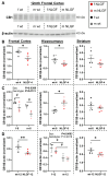
A) Representative blot of CB1 and β-actin. B) In 4-month AppNL-G-F mice, CB1 protein levels are significantly reduced in the frontal cortex and hippocampus, and trending down in the striatum. C) At 8-months, AppNL-G-F mice exhibit reduced frontal cortex CB1, compensation in the hippocampus, and no change in the striatum. D) At 12-months, CB1 is significantly reduced in the AppNL-G-F frontal cortex and striatum, but not the hippocampus. Sex differences were detected in the 8-month frontal cortex and 12-month hippocampus, with less CB1 protein in males in both cases. Normalized protein values are relative to exposure time and should not be compared between gels and graphs. Within each region, 4- and 12-month mice were on the same gels; see Figure S12 for age comparisons. Mean+/-SEM (n = 5-6/genotype/age). Abbreviations: CB1: cannabinoid receptor 1; kDa: kilodalton; m/f: male, female; NLGF: AppNL-G-F; wt: wild-type.
Figure 5
Figure 5
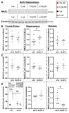
A) Representative blot of MAGL and β-actin. B) 4-month AppNL-G-F mice exhibit no differences in frontal cortical or striatal MAGL protein levels, with a trend to higher hippocampal MAGL levels. C) No genotype differences were detected at 8-months in any region. D) At 12- months, female AppNL-G-F mice exhibit increased MAGL protein levels. In the hippocampus, no changes were observed at 12-months. Striatal MAGL is reduced in 12-month AppNL-G-F mice. Normalized protein values are relative to exposure time and should not be compared between gels and graphs. Within each region, 4- and 12-month mice were on the same gels; see Figure S12 for age comparisons. Mean+/-SEM (n = 5-6/genotype/age). Abbreviations: gen: genotype; kDa: kilodalton; m/f: male, female; MAGL: monoacylglycerol lipase; NLGF: AppNL-G-F; wt: wild-type.
Figure 6
Figure 6
A) Representative summed [18F]MAGL-2102 PET images (0-60 min) and B) SUV curves demonstrating reduced MAGL availability following JZL184 treatment (MAGL inhibition) in 6-month wt and 5xFAD male mice (n = 3/genotype/treatment). 5xFAD mice exhibited higher MAGL availability than wt with or without JZL184. Mean+/-SEM. Abbreviations: ID/cc: injected dose per cubic centimetre; MAGL: monoacylglycerol lipase; PET: positron emission tomography; SUV: standardized uptake value; wt: wild-type.

References

    1. Varlow C, Boileau I, Wey H-Y, Liang SH, Vasdev N. Classics in Neuroimaging: Imaging the Endocannabinoid Pathway with PET. ACS Chem Neurosci. 2020;11:1855–62. - PMC - PubMed
    1. Hou L, Rong J, Haider A, Ogasawara D, Varlow C, Schafroth MA. et al. Positron Emission Tomography Imaging of the Endocannabinoid System: Opportunities and Challenges in Radiotracer Development. J Med Chem. 2021;64:123–49. - PMC - PubMed
    1. Lu H-C, Mackie K. An Introduction to the Endogenous Cannabinoid System. Biol Psychiatry. 2016;79:516–25. - PMC - PubMed
    1. Katona I, Freund TF. Endocannabinoid signaling as a synaptic circuit breaker in neurological disease. Nat Med. 2008;14:923–30. - PubMed
    1. Navarrete F, García-Gutiérrez MS, Jurado-Barba R, Rubio G, Gasparyan A, Austrich-Olivares A. et al. Endocannabinoid System Components as Potential Biomarkers in Psychiatry. Front Psychiatry. 2020;11:315. - PMC - PubMed

MeSH terms