. 2025 Feb 24;15(8):3424-3438.
doi: 10.7150/thno.103080. eCollection 2025.

Active microneedle patch equipped with spontaneous bubble generation for enhanced rheumatoid arthritis treatment

Affiliations

Active microneedle patch equipped with spontaneous bubble generation for enhanced rheumatoid arthritis treatment

Ting Liu et al. Theranostics. .

Abstract

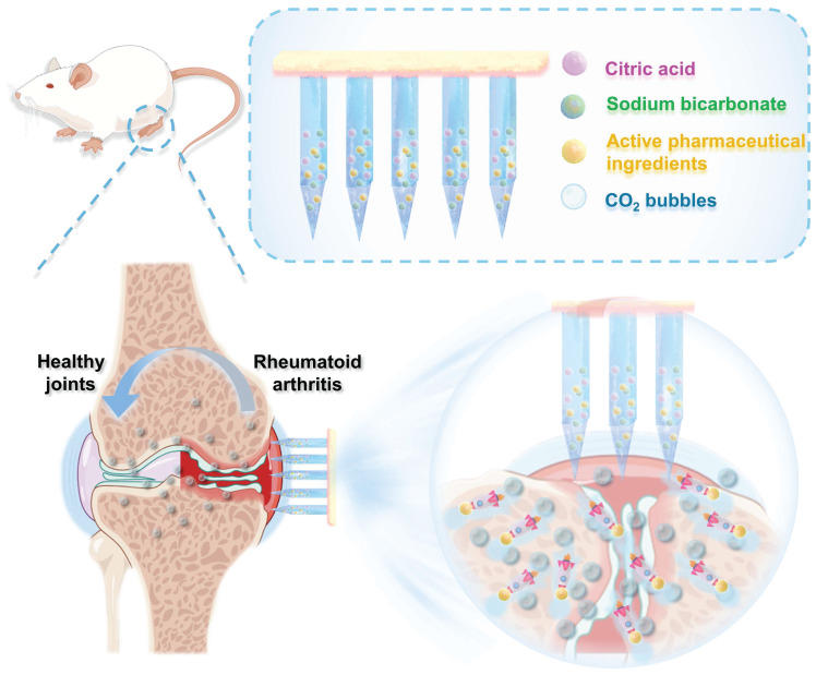
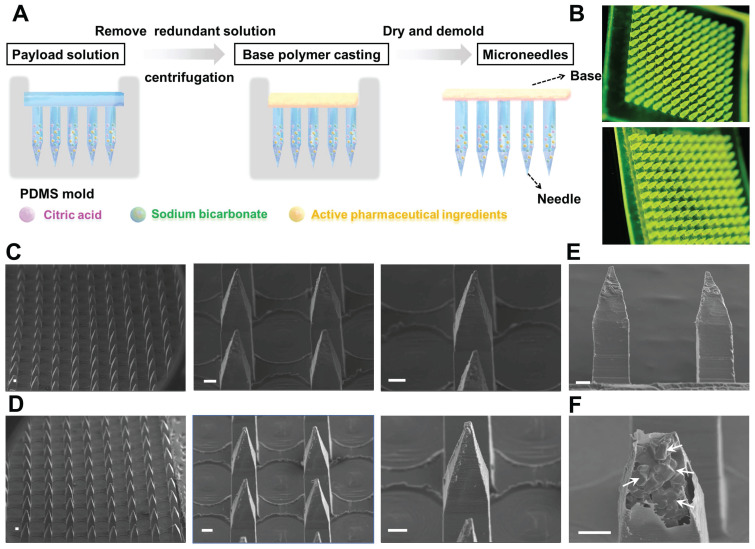
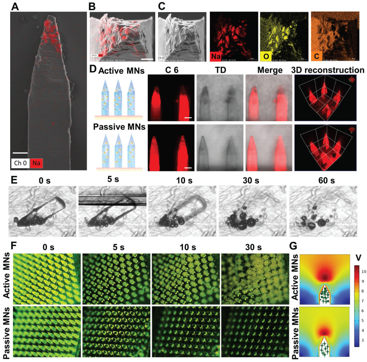
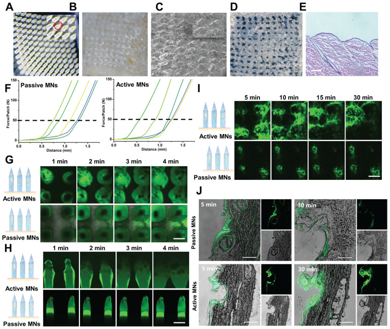
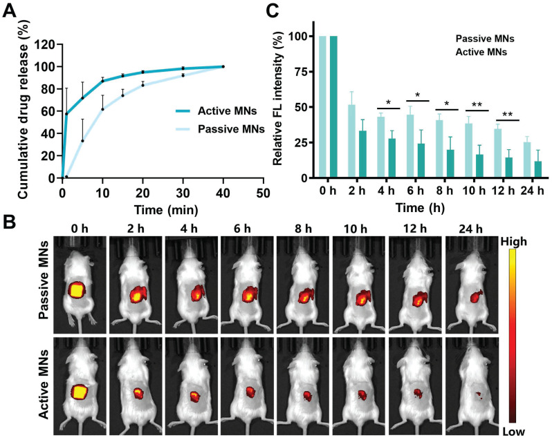
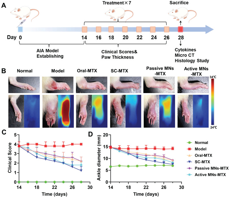
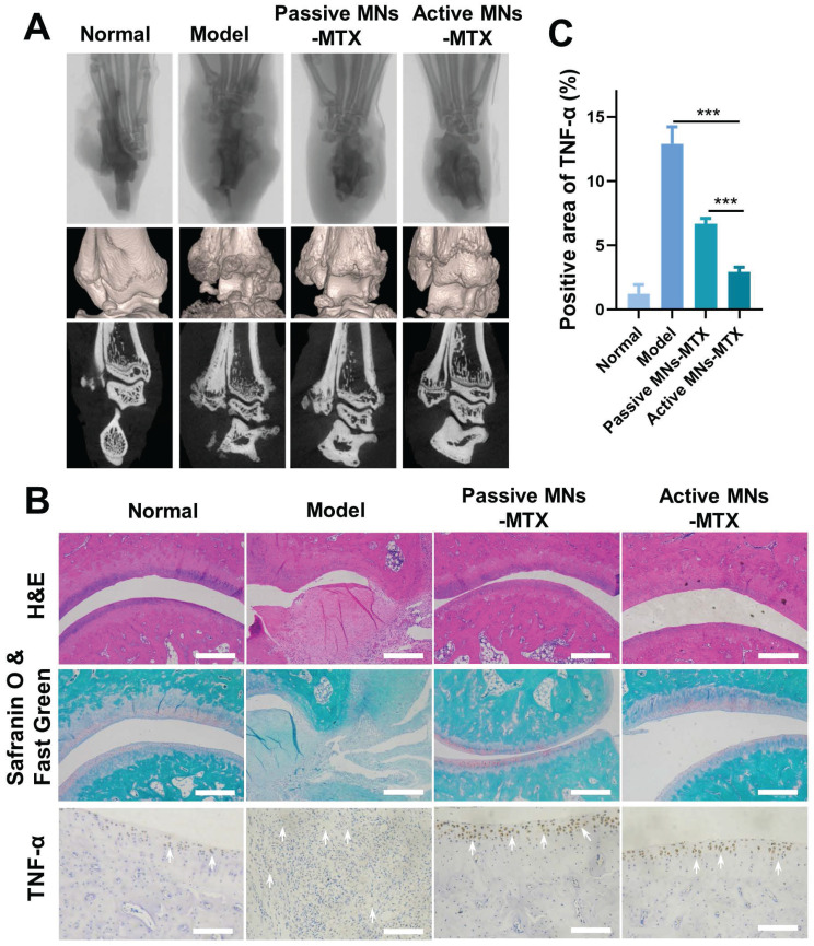
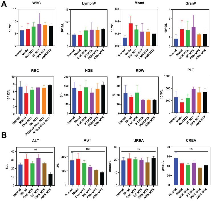
Rationale: The utilization of dissolving microneedles (MNs) facilitates the painless delivery of pharmaceuticals via the transdermal route. However, conventional MNs rely on passive diffusion through the gradual dissolving of the matrix, which can impede the therapeutic efficacy of the delivered drugs. Methods: In this study, we present the development of a novel degradable active MNs platform. This platform employs sodium bicarbonate and citric acid loaded in a dissolving MNs patch as a built-in motor for deeper and faster intradermal payload delivery. The sodium bicarbonate microparticles and citric acid undergo a chemical reaction when in contact with tissue fluid, resulting in the rapid formation of explosive carbon dioxide bubbles. This provides the necessary force to break through dermal barriers and enhance payload delivery. Results: The results demonstrated that the active MNs possessed excellent mechanical properties, rapid detachment characteristics, and superior drug release kinetics. Furthermore, the drug permeation behavior of active MNs exhibited enhanced permeation and distribution in skin-mimicking gel and porcine skin when compared to conventional passive MNs. In vivo experiments employing a rat model of rheumatoid arthritis showed that active MNs achieved superior therapeutic efficacy compared to passive MNs. Conclusions: This universal and effective autonomous dynamic microneedle delivery technology is straightforward to prepare and ultilize, and has the potential to improve the therapeutic efficacy of drugs, offering significant prospects for a diverse range of therapeutic applications.

Keywords: active microneedles; bubble generation; rapid separation; rheumatoid arthritis; transdermal drug delivery.


Conflict of interest statement

Competing Interests: The authors have declared that no competing interest exists.

Figures

Figure 1
Figure 1
Schematic illustration of the effervescent active MNs with spontaneous bubbles generation for enhanced drug delivery in RA treatment.
Figure 2
Figure 2
Preparation and characterizations of active MNs. (A) Schematic illustration of the preparation process of active MNs. (B) The optical handheld microscopy images of active MNs (upper) and passive MNs (lower) loaded with coumarin 6. (C, D) SEM images of active MNs (upper) and passive MNs (lower). (E) SEM image of the sideview of active MNs. (F) Magnified SEM image of inside active MNs (Scale bar = 100 μm).
Figure 3
Figure 3
The ingredient distribution and bubble generation behavior of MNs. EDX analysis of the side view (A) (Scale bar = 90 μm) and the top view (B) (Scale bar = 40 μm) of active MNs. (C) SEM images of single active MN tip and EDX analysis for sodium (Na), oxygen(O), and carbon (C). (D) CLSM and 3D reconstruction images of C6-loaded MNs (Red signal, scale bar = 200 μm). (E) Time-lapse images of single active MNs, showing bubble production and polymer dissolving in PBS. (F) Time-frame images of MNs, showing bubble production and drug diffusion in PBS. (G) COMSOL Multiphysics simulation of the flow generated by active MNs and passive MNs.
Figure 4
Figure 4
Mechanical strength and diffusion behavior of active MNs. (A, B) Images of active MNs before (A) and after (B) application to isolated rat skin. (C) SEM image of active MNs after application to isolated rat skin (Scale bar = 700 μm). (D) The photograph of the rat skin after active MNs insertion. (E) The H&E staining image of the rat skin sections after active MNs insertion (Scale bar = 50 μm). (F) The force curve of active MNs and passive MNs detected by the texture analyzer (n = 5). Time-lapse CLSM top view (G) and side view (H) images of active MNs and passive MNs inserted in gelatin-simulated skin, obtained at different time points (0-4 min). (I) Time-lapse CLSM top view images of porcine skin after application of active MNs and passive MNs at different time points (5-30 min). (Scale bar = 400 μm). (J) Fluorescence images of porcine skin cryosections after application of active MNs and passive MNs for 5 and 30 min. (Scale bar = 100 μm).
Figure 5
Figure 5
In vitro drug release and in vivo permeation profiles of MNs. (A) Cumulative in vitro release profiles of MTX from MNs. (B) In vivo fluorescence images of mice treated with active MNs and passive MNs and (C) its semiquantitative result.
Figure 6
Figure 6
Inhibitory effect of MTX-loaded active MNs on arthritic progression in AIA rats. (A) Schematic illustration of the treatment protocol for AIA rats. (B) Representative hind paw images of the normal, model, oral-MTX, SC-MTX, passive MNs-MTX, and active MNs-MTX groups. (C) Clinical scores of AIA rats in different groups. (D) Ankle diameter change of AIA rats in different groups. Data are shown as the mean ± SD (n = 5).
Figure 7
Figure 7
The effect of active MNs-MTX on relieving bone erosion, arthritic inflammation, and cartilage damage. (A) Representative images of Micro-CT imaging assessment. (B) Histological and immunohistochemical analysis of hind paws after various treatments. (Scale bar = 100 μm). (C) Semiquantitative analysis of inflammatory cytokine expression in joint tissues. Data are shown as the mean ± SD (n = 3).
Figure 8
Figure 8
Safety Evaluation. (A) Hematologic parameters of all experimental groups on day 16 (n = 3). (B) Serum biochemistry results of treated AIA rats (n = 3).

Similar articles

Cited by

References

    1. Liu Y, Xie W, Tang Z, Tan Z, He Y, Luo J. et al. A reconfigurable integrated smart device for real-time monitoring and synergistic treatment of rheumatoid arthritis. Sci Adv. 2024;10:eadj0604. - PMC - PubMed
    1. Hua P, Liang R, Yang S, Tu Y, Chen M. Microneedle-assisted dual delivery of PUMA gene and celastrol for synergistic therapy of rheumatoid arthritis through restoring synovial homeostasis. Bioact Mater. 2024;36:83–95. - PMC - PubMed
    1. Liu T, Fu J, Chen M, Wu Q, Quan G, Wu C. et al. In situ polymeric nanomicelle-generating dissolving microneedle patch for enhanced transdermal methotrexate delivery in rheumatoid arthritis treatment. Eur Polym J. 2024;210:113008.
    1. Xie J, Zhu X, Wang M, Liu C, Ling G, Zhang P. Dissolving microneedle-mediated transdermal delivery of flurbiprofen axetil-loaded pH-responsive liposomes for arthritis treatment. Chem Eng J. 2024;482:148840.
    1. Nooreen R, Nene S, Jain H, Prasannanjaneyulu V, Chitlangya P, Otavi S. et al. Polymer nanotherapeutics: a versatile platform for effective rheumatoid arthritis therapy. J Control Release. 2022;348:397–419. - PubMed