Review
. 2025 Feb 25;14(5):1539.
doi: 10.3390/jcm14051539.

Perspectives in the Diagnosis, Clinical Impact, and Management of the Vulnerable Plaque

Affiliations
Review

Perspectives in the Diagnosis, Clinical Impact, and Management of the Vulnerable Plaque

Alberto Alperi et al. J Clin Med. .

Abstract

Coronary artery disease is a highly prevalent disease that constitutes the leading cause of mortality worldwide. Acute coronary syndromes are the most devastating form of presentation of coronary disease, involving the acute formation of a thrombus within the coronary vessel lumen, further leading to flow limitation and diminished myocardial perfusion. Vulnerable plaques, which are characterized by thin-cap fibroatheroma, a large lipid pool, and macrophage infiltration and spotty calcification of the cap, pose a higher risk of coronary events despite not being flow-limiting. Iterations in intravascular imaging and coronary computed tomography have largely increased the ability to detect and define vulnerable plaques, and its clinical impact in early- and mid-term outcomes has been confirmed in several studies. In this review, we aimed to revise the current concept of vulnerable coronary plaque and its repercussion, to summarize the main pharmacological approaches for its management, and to provide an updated overview of the available evidence on preventive percutaneous interventional strategies in this clinical setting.

Keywords: coronary artery disease; percutaneous coronary intervention; vulnerable plaque.


Conflict of interest statement

The authors declare no conflict of interest.

Figures

Figure 1
Figure 1
Rates of major adverse cardiovascular events in the main studies evaluating the clinical impact of vulnerable plaques evaluated either with intravascular IVUS or OCT. IVUS: intravascular ultrasound. OCT: optical coherence tomography. Events were evaluated at 3-year follow-up for PROSPECT and VIVA trials, 1-year follow-up for ATHEROREMO and CLIMA trials, and 18-months follow-up for the COMBINEDFFROCT trial.
Figure 2
Figure 2
(Upper panel): Pathophysiology of atheroma plaque formation: 1. Endothelial dysfunction leads to increased LDL permeability and reduced nitric oxide production. 2. LDL molecules are oxidised in the subendothelial space (ROS: reactive oxygen species), and they promote the upregulation of adhesion molecules ICAM1 (intercellular adhesion molecule 1) and VCAM1 (vascular cell adhesion molecule 1). 3. Monocytes bind to these molecules and migrate into the vessel wall, where they convert into foam cells. 4. Excessive cholesterol uptake can disrupt autophagy regulation, induce cytotoxic effects, and ultimately lead to cell death. Additionally, inflammatory pathways—mediated by pro-inflammatory cytokines such as IL-18 and IL-6, as well as microRNAs—further drive the onset and progression of atherosclerosis. 5. Fatty streak is developed as an early stage of atherosclerosis, with no significant luminal narrowing. (Lower panel): Pathophysiology of vulnerable plaque development and progression: 1. Formation of necrotic core due to foam cell apoptosis and necrosis. 2. Thinning of fibrous cap: activated macrophages release matrix metalloproteinases and other enzymes that degrade extracellular matrix. 3. Neovascularization and intraplaque hemorrhage: immature microvessels rupture. 4. Plaque rupture and thrombosis: core exposure triggers thrombus formation.
Figure 3
Figure 3
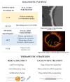
Diagnostic pathway and therapeutic strategies. Low attenuation plaque on CCT indicating vulnerability. On the OCT image the yellow arrows indicate the thin fibrous cap covering a lipid core extending 180° along the circumference (red dotted line). The white asterisk marks the wire artifact.

References

    1. Gaba P., Gersh B.J., Muller J., Narula J., Stone G.W. Evolving concepts of the vulnerable atherosclerotic plaque and the vulnerable patient: Implications for patient care and future research. Nat. Rev. Cardiol. 2023;20:181–196. doi: 10.1038/s41569-022-00769-8. - DOI - PubMed
    1. Virmani R., Burke A.P., Farb A., Kolodgie F.D. Pathology of the Vulnerable Plaque. J. Am. Coll. Cardiol. 2006;47:C13–C18. doi: 10.1016/j.jacc.2005.10.065. - DOI - PubMed
    1. Bentzon J.F., Otsuka F., Virmani R., Falk E. Mechanisms of plaque formation and rupture. Circ. Res. 2014;114:1852–1866. doi: 10.1161/CIRCRESAHA.114.302721. - DOI - PubMed
    1. Riley R.F., Patel M.P., Abbott J.D., Bangalore S., Brilakis E.S., Croce K.J., Doshi D., Kaul P., Kearney K.E., Kerrigan J.L., et al. SCAI Expert Consensus Statement on the Management of Calcified Coronary Lesions. J. Soc. Cardiovasc. Angiogr. Interv. 2024;3:101259. doi: 10.1016/j.jscai.2023.101259. - DOI - PMC - PubMed
    1. Virmani R., Kolodgie F.D., Burke A.P., Farb A., Schwartz S.M. Lessons from sudden coronary death: A comprehensive morphological classification scheme for atherosclerotic lesions. Arter. Thromb. Vasc. Biol. 2000;20:1262–1275. doi: 10.1161/01.ATV.20.5.1262. - DOI - PubMed

LinkOut - more resources