. 2025 Mar 17;21(3):e1012995.
doi: 10.1371/journal.ppat.1012995. eCollection 2025 Mar.

Eating the brain - A multidisciplinary study provides new insights into the mechanisms underlying the cytopathogenicity of Naegleria fowleri

Affiliations

Eating the brain - A multidisciplinary study provides new insights into the mechanisms underlying the cytopathogenicity of Naegleria fowleri

Ronald Malych et al. PLoS Pathog. .

Abstract

Naegleria fowleri, the causative agent of primary amoebic meningoencephalitis (PAM), requires increased research attention due to its high lethality and the potential for increased incidence as a result of global warming. The aim of this study was to investigate the interactions between N. fowleri and host cells in order to elucidate the mechanisms underlying the pathogenicity of this amoeba. A co-culture system comprising human fibrosarcoma cells was established to study both contact-dependent and contact-independent cytopathogenicity. Proteomic analyses of the amoebas exposed to human cell cultures or passaged through mouse brain were used to identify novel virulence factors. Our results indicate that actin dynamics, regulated by Arp2/3 and Src kinase, play a considerable role in ingestion of host cells by amoebae. We have identified three promising candidate virulence factors, namely lysozyme, cystatin and hemerythrin, which may be critical in facilitating N. fowleri evasion of host defenses, migration to the brain and induction of a lethal infection. Long-term co-culture secretome analysis revealed an increase in protease secretion, which enhances N. fowleri cytopathogenicity. Raman microspectroscopy revealed significant metabolic differences between axenic and brain-isolated amoebae, particularly in lipid storage and utilization. Taken together, our findings provide important new insights into the pathogenic mechanisms of N. fowleri and highlight potential targets for therapeutic intervention against PAM.


Conflict of interest statement

The authors have declared that no competing interests exist.

Figures
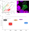
Fig 1
Fig 1. Naegleria fowleri trogocytosis is affected by inhibition of actin and tyrosine-protein kinase Src.
(A) Representative flow cytogram of CFSE-labeled Naegleria fowleri (green) in co-culture with tdTomato expressing human fibrosarcoma cells HT1080 (red). The degree of trogocytosis was determined by the number of Naegleria with ingested cell parts, represented by the red fluorescence of the tdTomato (orange) after 3 hours of incubation. (B) Live imaging of CFSE-labeled N. fowleri (green) and HT1080 (magenta) co-cultures showing the process of Naegleria adhesion to human cells. Ingested parts of HT1080 cells are visible in Naegleria vacuoles. Nuclei were labeled with Hoechst 33342 (blue). Scale bar =10µm. (C) Box-plot graph representing effect of Arp2/3 complex inhibitor (CK-666) and different types of kinase inhibitors (wortmannin, piceatannol and PP2) on the percentage of CFSE-labeled Naegleria with ingested tdTomato from HT1080 cells. Data were collected from four individual experiments. Statistical significance was determined using an ordinary ANOVA and Dunnett’s multiple comparison test. (** p-value<0.01, **** p-value<0.0001).
Fig 2
Fig 2. Proteomic changes in Naegleria fowleri co-cultured with bacterial or mammalian cells and amoebae isolated from infected mice.
(A) Venn diagram showing the overlap of upregulated proteins between short-term (6 h) Naegleria co-cultures with Klebsiella aerogenes or human fibrosarcoma cells HT1080, long-term (5 passages) co-culture with HT1080 cells and amoebae isolated from the brain of infected mice. (B) Venn diagram showing the overlap of 18 upregulated proteins between Naegleria long-term co-culture with human fibrosarcoma cells HT1080 and brain isolated amoebae. These proteins, listed in the table, include (marked in red) proteins involved in sulfur and lipid metabolism and lysozyme, which is discussed in the text as a potential novel virulence factor. Proteins were manually annotated by HHpred [70].
Fig 3
Fig 3. Predicted interactions between Naegleria fowleri (Nf) cystatin and cysteine proteases from Naegleria fowleri and human genomes.
(A) Best structures predicted by AlphaFold2 multimer from 69 N. fowleri cysteine proteases with their ipTM scores. (B) Best structures predicted by AlphaFold2 multimer from 237 Homo sapiens cysteine proteases, with their ipTM scores. N. fowleri and H. sapiens cysteine proteases are shown in blue. Nf cystatin is shown in orange, with its binding site in red.
Fig 4
Fig 4. Functional characterization of recombinant Naegleria fowleri (Nf) lysozyme, cystatin, and hemerythrin.
(A) Immunoblot detection of Nf lysozyme in cell lysates from axenically cultured N. fowleri (ax.) and amoebae co-cultured with Klebsiella aerogenes (g-), with Micrococcus lysodeikticus (g+), and with HT1080 fibrosarcoma cells (fib.) for 5 passages. Loading control is shown in S7B Fig. (B) Relative inhibition of human cathepsins K and L by recombinant Nf cystatin. The dotted line indicates 50% inhibition of peptidase activity (IC50). Relative inhibition values are related to enzyme activities with 0 nM Nf cystatin, which were taken as 100%. Enzymes were used at 50 nM concentrations. The IC50 values were extrapolated using non-linear regression (variable slope, 4 parameters). Bars represent S.D. (C) UV-vis spectra of recombinant Nf hemerythrin. Black line shows the deoxy form of hemerythrin obtained after reduction with sodium dithionite and methyl viologen under anaerobic conditions. The red line shows the oxidized form of hemerythrin with absorption maxima at 330 nm, 380 nm and 500 nm representing the O2-bound protein. The green line shows spectra after addition of 1 equivalent of nitric oxide (NO) to the fully reduced hemerythrin with a small band centered at 620 nm and other to absorption bands with maxima at 330 and 420 nm. (D) NO binding by hemerythrin measured by a modified Clark-type selective electrode showing four consecutive additions of NO stock solution to reach 20 μM, followed by the addition of 5 μM protein. (E) EPR spectra of as purified hemerythrin (red line), sodium dithionite and methyl viologen reduced hemerythrin (black line) and reduced hemerythrin incubated with 3 equivalents of nitric oxide (green line). Experimental conditions: protein concentration, 200 μM; temperature, 7 K; microwave frequency, 9.39 GHz; modulation amplitude, 1.0 mT; Microwave power, 2 mW. Spectral simulations and image preparation were performed using SpinCount [73].
Fig 5
Fig 5. Long-term co-culture of Naegleria fowleri with mammalian cells affects amoeba-secreted proteins.
(A) Axenic N. fowleri [65] and long-term co-cultured N. fowleri [32] proteins identified in their respective secretomes, sorted into 6 functional categories using Pfam motif identification and HHpred annotation. (B) Venn diagram showing the overlap of axenic N. fowleri and long-term co-cultured N. fowleri secretomes after incubation in an axenic medium. Four proteins identified only in the co-cultured amoeba secretome are listed in the table. (C) Volcano plot depicting significantly enriched proteins in the secretome of long-term co-cultured amoebae compared to the secretome of axenic amoebae. Both secretomes were prepared after one hour of incubation of amoebae with HT1080. (D) Graph showing changes in the percentage of CFSE-labeled Naegleria with ingested tdTomato from HT1080 cells after 1 and 3 hours of incubation of fibrosarcoma cells with either axenic amoebae or long-term co-cultured amoebae as measured by flow cytometry. (*** p-value<0.001).
Fig 6
Fig 6. Raman chemical maps of Naegleria fowleri grown axenically (A–D) and isolated from mouse brain (E–J).
A – bright field, B – merge, C – cytoplasm, D – nucleus, E – bright field, F – merge, G – cytoplasm, H – nucleus, I – ingested cells, J – lipid droplets, scalebar 5 µm; K – decomposed Raman spectra of axenic cell culture in the upper part and cells isolated from mouse brain in the lower part (shaded in orange). The intensities are normalized to the maximum value with an exception for the fingerprint region below 1800 cm−1 that has been doubled (marked as 2×) for better display of the spectra compared to the more intense high-wavenumber region above 2700 cm−1. However, the spectra of the ingested cells show an opposite trend, their spectrum has not been adjusted. Peak wavenumbers are displayed separately for four significantly different spectral clusters: cytoplasm (cyan), nucleus (blue), ingested cells (magenta), and lipid droplets (yellow). The characteristic peaks of amino acids and protein-related signals are highlighted by blue lines, nucleosides-, nucleotides- and nucleic acid-related components in yellow.

References

    1. Gharpure R, Gleason M, Salah Z, Blackstock AJ, Hess-Homeier D, Yoder JS, et al.. Geographic range of recreational water-associated primary amebic meningoencephalitis, United States, 1978–2018. Emerg Infect Dis. 2021;27(1):271–4. doi: 10.3201/eid2701.202119 - DOI - PMC - PubMed
    1. Dey R, Dlusskaya E, Oloroso M, Ashbolt NJ. First evidence of free-living Naegleria species in recreational lakes of Alberta, Canada. J Water Health. 2023;21(3):439–42. doi: 10.2166/wh.2023.325 - DOI - PubMed
    1. Martínez-Castillo M, Cárdenas-Guerra RE, Arroyo R, Debnath A, Rodríguez MA, Sabanero M, et al.. Nf-GH, a glycosidase secreted by Naegleria fowleri, causes mucin degradation: an in vitro and in vivo study. Future Microbiol. 2017;12(9):781–99. doi: 10.2217/fmb-2016-0230 - DOI - PMC - PubMed
    1. Cervantes-Sandoval I, Jesús Serrano-Luna J, Pacheco-Yépez J, Silva-Olivares A, Tsutsumi V, Shibayama M. Differences between Naegleria fowleri and Naegleria gruberi in expression of mannose and fucose glycoconjugates. Parasitol Res. 2010;106(3):695–701. doi: 10.1007/s00436-010-1727-z - DOI - PubMed
    1. Carrasco-Yepez M, Campos-Rodriguez R, Godinez-Victoria M, Rodriguez-Monroy MA, Jarillo-Luna A, Bonilla-Lemus P, et al.. Naegleria fowleri glycoconjugates with residues of α-D-mannose are involved in adherence of trophozoites to mouse nasal mucosa. Parasitol Res. 2013;112(10):3615–25. doi: 10.1007/s00436-013-3549-2 - DOI - PubMed

MeSH terms

LinkOut - more resources